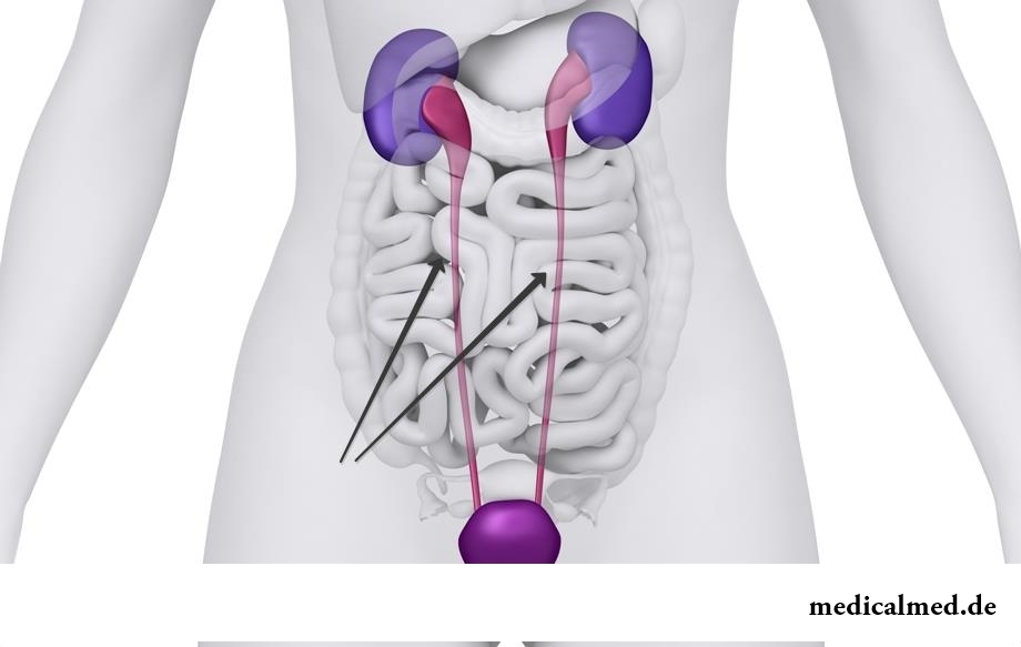
Ureter
Ureter – pair urinary body which serves for removal of urine in a bladder.
Ureter structure
The ureter begins from the narrowed site of a renal pelvis where the urine formed in a kidney flows down. Its output end comes to an end in a bladder wall. In this place mucous forms the fold interfering the return current of urine. The fold works as the valve as thanks to the muscle fibers which are contained in it it can actively be closed.
Externally the ureter has an appearance of a thin tube which has an outside cover from connecting fabric, an average muscular layer which fibers intertwine diversely, and an internal mucous membrane which forms longitudinal folds on all length of an ureter.
Part of an ureter is located in an abdominal cavity, and a part – in a cavity of a small pelvis. On all length its pieces of narrowing alternate with expansions. Diameter of this body in an abdominal cavity averages from 8 to 15 mm, in a small basin – to 6 mm. Considerable elasticity allows to extend to an ureter at difficulty of outflow of urine to 8 cm, for example, if in an ureter stones. The bottleneck – an exit from a renal pelvis, and it is biologically reasonable.
Functions of an ureter
The main function of an ureter is timely removal of the urine formed in kidneys in a bladder. At first the upper part of an ureter is filled, and thanks to reductions of muscle fibers in its wall urine moves ahead further, in a bladder, even in horizontal position of the person.
Inspection of ureters
Begin inspection with collecting complaints. Most often patients with diseases of ureters complain of a pain syndrome. Pains can be pricking, aching, pristupoobrazny, to give down a stomach. Defeat of pelvic department can give disturbance of a rhythm of an urination – a dysuria.
At a palpation of a stomach there can be tension of front its wall and morbidity on the ureter course. The lower piece of this body manages to be probed at a research through a vagina at women or a rectum at men.
In analyses of urine at pathology of ureters leukocytes and erythrocytes can be found. Most often this evidence of inflammatory changes or stones in ureters.
Tsistoskopiya allows to examine mouths of ureters in a bladder – their form, the sizes, an arrangement, availability in them of blood or purulent discharges. Hromotsistoskopiya allows to define the block of advance of urine because of a stone in an ureter or damages. Damage level can be determined at catheterization more precisely, besides it can become remedy for an ureter in need of removal of urine.
At survey urography ureters are not visible, but it is possible to notice in them X-ray contrast concrements. Their course is visible at a research with contrast – excretory urography. In these cases it is possible to find asymptomatic doubling of ureters also. In case of introduction of contrast from a bladder cavity the research is called a retrograde ureterografiya.
Sokratitelny opportunities investigate using X-ray cinematography, an elektroureterografiya. These types of inspections allow to reveal such dysfunctions of an ureter, hypo - or a hyperkinesia, hyper - or an atony.
Diseases of an ureter and approaches to their treatment
Distinguish the inborn and acquired pathology of ureters. Inborn diseases arise under the influence of disturbing factors on a fruit.
The hypoplasia often happens at an underdevelopment of the corresponding kidney. Diameter of an ureter decreases, in some places it can obliterate. Narrowing, or a stenosis, is most often formed in a vesicoureteral segment. In these cases perhaps operational treatment of an ureter with plastics of the struck segment.
Valves of an ureter are a doubling of an internal cover of body in the form of a fold, they meet quite seldom.
Inborn atony – one of the heaviest pathologies. Due to the lack of reductions the ureter very strongly extends. Clinically it can not be shown in any way, but in urine the resistant pyuria is found.
Acquired diseases generally are a consequence of disturbance of passability. It can be a consequence of a prelum from the outside or existence of an obstacle in a gleam.
From the outside of a prelum long locks, ureter excesses, gynecologic pathology, cancer diseases of a bladder, prostate, neck of uterus most often make.
At an urolithiasis small stones from a pelvis of a kidney can get to an ureter, breaking urine outflow. Surgery an ureterolithotomy is made for the purpose of removal from an ureter of stones if other methods were inefficient.
The cancer tumor, chronic inflammatory process (for example can cause obstruction of body except stones, at tuberculosis, a schistosomiasis). Treatment of ureters will consist in elimination of an obstacle or removal of an ureter in the operational way and drainage of a renal pelvis.
At fibrous defeat in area of retroperitoneal cellulose there is a fibrous polyureteritis. The ureter in this case is captured outside by fibrous fabric in the form of the coupling which squeezes it outside. This pathology can also be corrected only in the surgical way.
There are very curious medical syndromes, for example, persuasive swallowing objects. In a stomach of one patient suffering from this mania 2500 foreign objects were revealed.

What they, women? Beautiful, gentle, passionate and at the same time windy, gusty, and nervous. And what is stranger: all эт...
Section: Articles about health
All parents are ready to what the baby often and pisat much. Since then, as the absorbing diapers strongly became current, keeping of the kid in dryness does not represent any problems. But if the grown-up kid continues to urinate in panties, parents of a nacha...
Section: Articles about health
During foot walks blood moves on vessels more actively and one and all bodies are supplied with a large amount of oxygen. It affects the state of health of the person very positively....
Section: Slideshow
Urogenital candidiasis (milkwoman) – a fungal infection which annoys unpleasant feelings in the field of generative organs, сопр...
Section: Articles about health
Tuberculosis – a serious infectious disease which development is caused by mycobacteria (Koch's bacilli). The illness is known from an extreme antiquity. Long time fight against it was considered as ineffective. Quite often the disease affected the whole families, and mortality from it was very much...
Section: Articles about health
The stroke is one of the most widespread diseases of the person, annually in the world about 6 million cases of this pathology are registered. According to medical statistics, strokes occur almost three times more often than myocardial infarctions. The disease belongs to heavy, and has an unfavourable result: the lethality reaches 40% among women and 25% among men. A considerable part of the patients who endured a stroke cannot be recovered completely. We suggest readers to examine...
Section: Articles about health
To look healthy and means well-groomed not only to be pleasant to people around, but also to feel strong, sure and taken place. To Spa...
Section: Articles about health
On the head of the person about one million hair follicles, or as they are called still, hair bulbs are located. At the time of the birth most of them is in the "sleeping" state, but within several weeks follicles become more active, and from them begin р...
Section: Articles about health
Aging — natural and inevitable process. Over time our skin loses elasticity, on it saggings are formed, the face form loses former clearness. The procedure of nitevy lifting (nitevy tightening) can successfully solve this problem. In order that it is better to get acquainted with this popular procedure, we will tell you 6 cognitive facts about it....
Section: Articles about health
Season of activity of viral infections in the heat. Everyone can get sick, but probability of this unpleasant event it is possible and it is necessary miny...
Section: Articles about health
The summer of this year in Russia was very ambiguous. Regions suffered from a merciless heat, from pouring rains, the hail from time to time dropped out, then there was again a heat which alternated with rainfall again. Many people suffer from such sharp changes of weather...
Section: Articles about health
Healthy lifestyle today in fashion, and many parents think of that the child from the early childhood played sports. Trainings will help it to become strong and hardy, will improve coordination of movements, and also will exert positive impact on mentality: it will become more collected and purposeful....
Section: Articles about health
Feeding by a breast - the integral part of ideal motherhood allowing to come into contact with the kid and to create since early years...
Section: Articles about health
They say that to ensure health and longevity of people it is obliged. Really, at competent approach to these questions, minimization of an adverse effect of many factors does not represent a special problem. Practically everyone has an opportunity to play sports...
Section: Articles about health
Summer in the heat. Many are going to spend vacation abroad. Travelers the tender seas, rest on beaches wait, for sightseeing, campaigns on natural and cultural reserves. But, unfortunately, on vacation also problems with health can wait for us. On a foreign trip it is possible to face also diseases which not only will spoil long-awaited issue, but also will force to be treated within long months after its termination. To be insured completely from troubles of it a sort...
Section: Articles about health
The word "onikhokriptoz" is unfamiliar to most of people, meanwhile quite so physicians call very widespread problem: growing...
Section: Articles about health
Diapers for adults – individual one-time means of hygiene which in some situations is irreplaceable and from such situations any person is not insured. Though nobody perceives need of their use with enthusiasm, however without it to a sra...
Section: Articles about health
High temperature - a frequent symptom of such widespread diseases as a SARS, quinsy, pneumonia, etc. To reduce heat, having facilitated a condition of the patient, doctors recommend to accept antipyretics, however their use is not always possible. Too frequent use of these drugs can lead to allergic reactions, and also overdose, causing poisoning. It happens also that there are no antipyretics simply in the house. In these situations it is pertinent to use it...
Section: Articles about health
On health of the person physicians know about salutary action of animals long ago. About 7 thousand years ago great Hippocrates рекоменд...
Section: Articles about health
The pine is one of the most widespread plants of our woods. Its needles and pitch not without reason called by "gallipot" were since ancient times used for strengthening of protective forces of an organism, treatment of avitaminosis, anemia and many other diseases. In recent years wide п...
Section: Articles about health
The hysteromyoma is diagnosed more than at a third of women 35 years are more senior. This high-quality new growth which at early stages successfully resolves by means of medicines. It is necessary to resort to an operative measure only when patients too late address specialists, or therapeutic methods do not give the expected effect because of specific features of an organism. Besides, there is a large number of quite effective national...
Section: Articles about health
Within several decades of our compatriots convinced that the use of butter nasty affects on...
Section: Articles about health
Some people consider what for medicine of the 21st century of secrets in the field of health of the person almost does not exist. It absolutely not so. The more answers scientists receive, the more the most difficult questions are raised for them by life. Besides, there are diseases, not объясн in any way...
Section: Articles about health
We present to yours the TOP of the medicamentous means exerting the stimulating impact on a potentiality, i.e. on ability of the man to commission of sexual intercourse. At once it is necessary to tell that not always disturbances of erectile function can be eliminated with reception of this or that drug. The reasons of decrease in a potentiality there can be a set, from banal overfatigue before tumoral process in a small basin therefore if the man faces similar problems too often, it should turn...
Section: Articles about health
Women quite often suffer from complexes concerning the sizes of the bust. Strangely enough, reason душевног...
Section: Articles about health
The majority of gynecologic diseases prove three main signs, each of which speaks about need of a visit to the gynecologist. Certainly, it is possible to establish the exact diagnosis only after inspection, but on the basis of some signs it is possible пр...
Section: Articles about health
Reactive pancreatitis - the disease which is characterized by inflammatory process in a pancreas which arises most often because of excess activity of digestive enzymes. It − the emergency state which treatment has to take place in surgical department under control of doctors. The acute inflammation of gland can become the reason of its transition to a chronic form, and also development is purulent - necrotic pancreatitis which the extensive necrosis of fabrics can follow. Zabolev...
Section: Articles about health