X-ray analysis
What is represented by a X-ray analysis
 The X-ray analysis represents a research of internals and systems of a human body by their projection to special paper or a film by means of X-ray. The X-ray analysis is the first method of medical visualization which allowed to receive the image of fabrics and bodies, to investigate them at human life. This diagnostic method was open in 1895 when the German physicist Wilhelm Konrad Rentgen registered property of x-ray emission to darken a photographic plate.
Because the X-ray analysis assumes a research of three-dimensional objects which on a X-ray sensitive film are represented by flat, it is necessary to do pictures at least in two projections to find localization of the pathological center.
It is possible to carry the following to advantages of a X-ray analysis:
- ease of carrying out and wide availability;
- lack of special preparation for the majority of researches;
- rather low cost, except for researches with obtaining results in a digital form;
- absence the operator dependence that allows to use the obtained data on consultation at various specialists.
Despite wide circulation, the X-ray analysis has also the shortcomings:
- the image turns out "frozen" that complicates assessment of function of body;
- harmful effects of ionizing radiation on the studied organism;
- low informational content in comparison with modern tomographic methods that is explained by projective stratification of anatomical structures the x-ray image;
- need of use of the contrasting substances at a X-ray analysis of soft tissues.
Thorax X-ray analysis
This diagnostic method allows to investigate lymph nodes, blood vessels, respiratory tracts, lungs, heart. Usually the X-ray analysis of a thorax assumes two pictures, from a breast and from a back, however in case of serious condition of the patient also one picture is admissible. Before this research no special preparation is required, however because of a negative impact of radiation on a fruit it is not recommended to carry out a X-ray analysis during pregnancy.
The X-ray analysis of a thorax is appointed in the following cases:
- for definition of the reason of cough, an asthma or a stethalgia;
- at such cardiological problems as heart failure or the increased heart;
- for the purpose of diagnosis of lung cancer, pneumonia, a chronic obstructive pulmonary disease, a mucoviscidosis, pheumothorax;
- for detection of fractures of edges, small damages, and also the problems causing pulmonary hypostasis;
- for the purpose of identification of foreign objects in lungs, airways and a stomach.
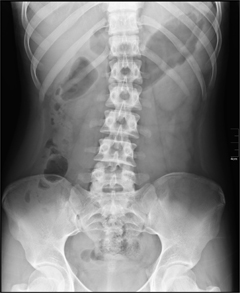
Backbone X-ray analysis
The pictures received as a result of a backbone X-ray analysis allow to define a structure and density of a bone tissue, shift of vertebrae, existence of erosion, to reveal roughness of contours and sites of thinning or a thickening of a bast layer of bones. It is reasonable to conduct this research in the following cases:
- for diagnosis of deformation, incomplete dislocations, changes and shift of vertebrae;
- for the purpose of identification of degenerative changes of a backbone, infectious diseases and inborn malformations;
- for assessment of a condition of a backbone at metabolic frustration and arthritis;
- for the purpose of identification of defeats of intervertebral disks.
The X-ray analysis of a backbone does not demand any special preparation, it is necessary to follow strictly only during the research to instructions of the doctor, adopting the necessary provision on a x-ray table and holding at some point the breath.
X-ray analysis of lungs
 The doctor can appoint a X-ray analysis of lungs in the presence at the patient of such symptoms as a pneumorrhagia, dry cough, the general weakness, the increased temperature, loss of weight, pain in lungs or to back. This research allows to diagnose tuberculosis, pneumonia, tumors or fungus diseases of lungs, and also to reveal foreign bodys.
As a rule, the X-ray analysis of lungs assumes receiving two pictures – by means of front and side roentgenograms. Small children during this research should be in lying situation, and the doctor at assessment of the roentgenogram has to consider the changed proportions and features of blood supply of lungs when finding the person in horizontal position on spin. The X-ray analysis of lungs does not assume any special preparation.
X-ray analysis of joints
This diagnostic method is used usually at chronic or long arthritis, and also at suspicion on the deforming osteoarthrosis. At other rheumatic diseases in most cases the X-ray analysis of joints allows to reveal symptoms much later, than laboratory researches or observations of the overall clinical picture. However x-ray films are all the same necessary, they will allow to compare results of further researches to initial data.
In case of a research of symmetric joints the roentgenogram becomes in direct and side projections, and at diagnosis of diseases of shoulder or hip joints one more additional projection – slanting is required.
For detection of a disease data of a X-ray analysis of joints are analyzed in the following order:
- the outline of a joint crack – its narrowing testifies to an initial stage of a pseudorheumatism;
- the joint ends of bones - their bone structure, a ratio, a form, the sizes;
- condition of periartikulyarny soft tissues;
- contours of a cortical layer.
At assessment of a X-ray analysis of joints the clinical picture, prescription of a disease and age of the patient is considered.
Skull X-ray analysis
Surprisingly, but this method малоинформативен at diagnosis of craniocereberal injuries, but at the same time the X-ray analysis of a skull is reasonable in the following cases:
- for the purpose of detection of fractures of skull;
- for diagnosis of a tumor of a hypophysis;
- at detection of inborn malformations;
- for diagnosis of some metabolic and endocrine diseases.
The doctor can direct to a skull X-ray analysis in the presence of such symptoms as a loss of consciousness, dizziness, head pains, disturbance of a hormonal background.
As a rule, this research is carried out in five projections – left and right side, perednezadny, posteroanterior and axial. The X-ray analysis of a skull does not assume any special preparations, the only requirement – lack of any metal objects (jewelry, dentures, points) in a radiation zone.
Except the listed types of a X-ray analysis, the stomach and a duodenum, a gall bladder and biliary tract, a large intestine, various departments of a peripheral skeleton, an abdominal cavity, a cavity of the uterus and passability of fallopian pipes, and also teeth can be investigated by the same way.
The weight of a human brain makes about 2% of all body weight, however it consumes about 20% of the oxygen coming to blood. This fact does a human brain extremely susceptible to the damages caused by shortage of oxygen.

History of mankind contains several tens of epidemics whose emergence was compared by eyewitnesses and historians to doomsday. With...
Section: Articles about health
According to data of World Health Organization, the cataract is diagnosed almost for 7% of the population of Earth. The statistics of incidence is considered not full as at an initial stage the illness, as a rule, does not cause to the person of special inconveniences, and many having got sick...
Section: Articles about health
The list of stereotypes of which, apparently, all know strongly includes following: British surely eat porridge for breakfast. Perhaps, not all modern residents of Britain arrive quite so, but for those from them which continue to follow this tradition, it is possible to be glad sincerely: oat flakes are a product which regular use not only helps the person to keep force and beauty long. Porridge in a special way influences an organism, protecting it from seriousness...
Section: Articles about health
Dietary supplements (dietary supplements) for the last decades were so thoroughly included into our life that, apparently, it is already impossible on...
Section: Articles about health
Olive oil – the product capable to make a powerful contribution to health of the person if it includes it in the diet. The rich vitamin composition of oil does it by a product number one from many diseases including from deadly. Only two tablespoons...
Section: Articles about health
The brain of the person is studied not one hundred years, but the quantity of the riddles connected with this body increases rather, than decreases. Perhaps, numerous delusions concerning a structure and functioning of a brain are explained by it, many of which arose long ago, but continue to exist and today. Such we are ready to acquaint readers with the most widespread myths....
Section: Articles about health
Tick-borne encephalitis – one of the most dangerous viral diseases which causative agents transfer and is given to people by ixodic mites. Эт...
Section: Articles about health
There is a lot of fans of beer in our country. Statistically, on each average Russian (including women and children) in a year about 60 liters of this drink are consumed. It is not a lot of, as in the Czech Republic or Germany, but figure all the same impressive. Radova...
Section: Articles about health
Deciding to get rid of an addiction, not all imagine what effects it is necessary to face. Process of refusal of smoking causes quite essential discomfort in most of people: differences of mood, a sleep disorder, fatigue, decrease in physical and intellectual activity and a number of other symptoms reducing quality of life. Abstinence can be strong: an essential part of attempts comes to an end leaving off smoking failure, and people are returned to the use of cigars...
Section: Articles about health
Radiological methods of a research are applied in medicine more than hundred years, and thanks to them millions of lives were saved. In m...
Section: Articles about health
The healthy nutrition is the invariable principle of health and good health for long years of the woman. Nevertheless, in a diet at each stage of life there are the features allowing to support an organism by those substances which are most necessary...
Section: Articles about health
Ayurveda - the most ancient tselitelsky practice which came to us from India. It represents the doctrine about maintenance of physical, psychological and moral health of the person by means of the complex of procedures including a diet, cleaning of an organism, breathing exercises, massage, and in case of a disease - and medicinal therapy. The healers practicing Ayurveda assign very important part to spices, and at the heart of Ayurvedic drugs, as a rule, there are they. It is considered that spices not of t...
Section: Articles about health
It is possible to find the extensive range of fruit and vegetables in modern shops. Russians already got used that on counters in any...
Section: Articles about health
For anybody not a secret that our country is one of the most "drinking" in the world. At clear understanding that the use of hard alcoholic drinks – occupation extremely harmful, most of Russians belong to alcoholism with unjustified loyalty. These...
Section: Articles about health
Tuberculosis – a serious infectious disease which development is caused by mycobacteria (Koch's bacilli). The illness is known from an extreme antiquity. Long time fight against it was considered as ineffective. Quite often the disease affected the whole families, and mortality from it was very high. It became the reason of emergence of a set of delusions concerning transmissibility and a possibility of treatment of tuberculosis....
Section: Articles about health
The thought that the mass of their body is too big at least once in life visits from 80 to 95% of women. Many...
Section: Articles about health
We live during an advertizing era. Daily each person receives a solid portion of persuasive councils about what to eat to be healthy and successful. Products about which we will talk today are combined by the following circumstance: all of them are positioned as most...
Section: Articles about health
So, you resolved to lose weight. And now you try to understand what to begin with: from exercise stresses or a diet? And how to make that process of weight loss did not give you an inconvenience, and, on the contrary, brought joy?...
Section: Slideshow
The person, as well as all other beings living on our planet feels weather changing. It is normal meteosensitivity, not...
Section: Articles about health
White teeth and the Hollywood smile – a dream of many people. Long time was considered that the plaque on teeth and change of their color – destiny of those who incorrectly eat smokes and badly brushes teeth. But the paradox is available: at everything the variety of toothpastes existing today...
Section: Articles about health
Stroke (acute disorder of cerebral circulation) – one of the most widespread neurologic diseases. Annually in the world more than 6 million people die of this illness. From the survived patients about 80% become disabled people, and nearly a third from them needs afterwards permanent care. In fact, the stroke creates a situation at which a part of cells of a brain loses blood access, loses an opportunity to receive oxygen and nutrients, and perishes. As a result of a razviv...
Section: Articles about health
The word "onikhokriptoz" is unfamiliar to most of people, meanwhile quite so physicians call very widespread problem: growing...
Section: Articles about health
For the last decades the diabetes mellitus of the second type became really world problem. The number of cases annually increases, and average age of patients for whom the illness is diagnosed, steadily decreases. Specialists consider that one of osno...
Section: Articles about health
Within several decades of our compatriots convinced that the use of butter nasty affects a condition of coronary vessels. As a result the reputation of a product was impaired thoroughly a little, and many almost ceased to include it in the diet, having given preference "to safer" to vegetable fats. Meanwhile, the last researches showed that harm of butter for health is strongly exaggerated. But the product has a number of unique properties, to...
Section: Articles about health
The pine is one of the most widespread plants of our woods. Its needles and pitch not without reason called by "gallipot", since ancient times испол...
Section: Articles about health
The phenomenon of the panic attack is known long ago, but the reasons of its emergence still are up to the end not found out. It is established that more than 30% of people at least once in life become the victims of very unpleasant phenomenon: without everyones on that the reasons they have a feeling of horror, with...
Section: Articles about health
For the time being the perspective of heart diseases seems to most of people remote and foggy. But sooner or later practically each adult faces extremely unpleasant feelings: sudden stethalgia. To be consoled at this time in a thought of what for a heart attack still early, will hardly turn out: if the person is impressionable, he, as a rule, has feeling of panic and fear of fast death. And meanwhile, it is very often possible to confuse pains with cardiac pains невралгическог...
Section: Articles about health