

Ultrasonography of a uterus
 Ultrasonography of a uterus and appendages – the ultrasonic inspection giving an idea to the gynecologist of a condition of a uterus and appendages (uterine tubes, ovaries), their form, the size, structure.
By results of ultrasonography often it is possible to draw a conclusion on the infertility reason, to diagnose a gynecologic disease.
Features of performing ultrasonography of a uterus and appendages
Ultrasonography of a uterus there undergo women in whom at standard inspection and on the basis of complaints symptoms of a gynecologic disease were found: pains in a stomach bottom to or during periods, long and painful periods, intermenstrual bleedings.
Besides, carry out ultrasonography of a uterus at pregnancy. Inspection is appointed for confirmation of the fact uterine (or extrauterine) by pregnancies, exceptions of a vesical drift. Ultrasonography of a uterus at pregnancy on early term helps to find out a condition of pregnancy, its term, and in the presence of pathologies – an essence of a problem and a possibility of purpose of treatment or need of abortion,
High-quality examination can be conducted only at the filled bladder therefore the woman several hours prior to ultrasonography is recommended to drink 1,5-2l waters. Carry out ultrasonography for 5-7 day of a cycle.
Ultrasonography of appendages is appointed to pass to women who have complaints to irregular menstrual bleedings and their absence, pains in a stomach bottom, infertility.
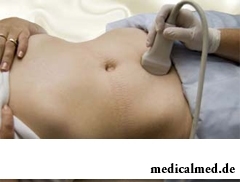
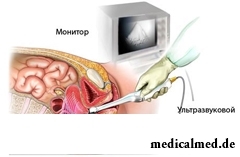
As well as at ultrasonography of a uterus, at inspection of pipes and ovaries if examination is conducted outside, through a front wall of a peritoneum, the woman needs to fill a bladder. If to the patient appointed to undergo transvaginal inspection (the sensor is entered inside into a vagina), specially it is not necessary to prepare and fill a bladder.
Ultrasonography of appendages is desirable to take place in the same terms, as ultrasonography of a uterus. If it is necessary to study development of follicles of ovaries, ultrasonography is carried out several times throughout a menstrual cycle.
Results of ultrasonography of a uterus
Results of inspection – both abdominal, and transvaginal, the woman receives on a hand soon after ultrasonography, on the same day.
 In the conclusion of the doctor information on a shape of a uterus, its size, thickness of walls and an endometria, compliance to its day of a cycle, existence of deviations in development, structure of a neck of uterus, on the sizes of ovaries, their structure, existence of the ripening follicles moves. Data on uterine tubes in results of ultrasonography of the healthy woman should not be - pipes without pathologies on ultrasonography are not visible.
Norms for the sizes of a uterus of the nonpregnant woman: length – 70 mm, width – 60 mm, the perednezadny size – 42 mm.
Norms of the sizes for ovaries: width – 25 mm, length – 30 mm, thickness – 15 mm.
Besides in results of ultrasonography of a uterus there can be data on the found diseases:
- myoma – a tumor of a muscular layer (myometrium) of a uterus of high-quality character. Ultrasonography can reveal myoma in only 1 cm in the diameter;
- endometriosis. A disease at which the cover of a uterus goes beyond a cavity. By means of ultrasonography it is impossible to make the final diagnosis since only small bubbles in a cavity of the uterus are visible, but inspection gives the chance to appoint additional analyses;
- polyps. Arise when the mucous membrane of a uterus expands. On ultrasonography the doctor at this disease notices a thickening or growth of an endometria;
- malformations. So call manifest deviations in sizes, a form, a uterus location. Ultrasonography of a uterus gives the chance to diagnose a two-horned, underdeveloped, saddle uterus. If the malformation is found, to the woman enter contrast for more detailed studying of body;
- cancer of a neck or endometria. The new growth of the malignant nature found on a neck of uterus or in its mucous;
- polycystosis of ovaries. The disease arising because of a hormonal imbalance. On ultrasonography the doctor notes a thickening of capsules of ovaries, increase in ovaries, finds cysts;
- salpingitis. An inflammation of uterine tubes because of which in them the commissures capable to become the infertility reason are formed. On ultrasonography it is visible that uterine tubes are thickened.
- tumors of pipes and ovaries. Ultrasonography of appendages gives the chance to find tumors, to estimate their size and structure. For more exact diagnosis and purpose of treatment the patient needs to undergo additional inspection.
Caries is the most widespread infectious disease in the world to which even flu cannot compete.

There is an opinion that at low temperatures safety of products is ensured longer and better thanks to what the refrigerator considers...
Section: Articles about health
Many of us, probably, noticed more than once that from intellectual loadings at some point the brain as though "overheats" and "assimilation" of information is strongly slowed down. Especially this problem urgent for persons of age becomes more senior than fifty years. "It is already bad with...
Section: Articles about health
Good appetite was always considered as a sign of good health. The correct operation of the mechanism which is responsible for the need for nutrients and receiving pleasure from process of its satisfaction demonstrates that the organism functions without special deviations. On the other hand, appetite of the person is not a constant. It depends on the culture of food, flavoring addictions imparted since the childhood which can change during life, weather, mood and many д more than once...
Section: Articles about health
It is impossible to imagine human life in which there would be no plants. Practically in each apartment and any of productions...
Section: Articles about health
The metabolism at each person proceeds in own way. However between the speed of this process and disposal of excess weight after all all have a dependence. Unfortunately, the people inclined to try on itself numerous "miracle" diets, not always at...
Section: Articles about health
Wood louse – the ordinary-looking unpretentious plant extended in all territory of our country. It quickly expands, and sometimes fills sites, bringing a lot of chagrin to gardeners. Perhaps, they would be upset less if knew that the wood louse is valuable medicinal raw materials. A, C and E vitamins, organic acids, tannins, wax, saponins, lipids, mineral salts and essential oils are its part....
Section: Articles about health
The number of long-livers is very small. One person from 5 thousand lives up to age of 90 years, and the centenary boundary steps only about...
Section: Articles about health
Energy saving lamps are one of the most popular products of innovative technologies, and there is no wonder: they much more economic also are more long-lasting than usual filament lamps. At the same time there are fears that energy saving bulbs can become the reasons...
Section: Articles about health
On health of the person physicians know about salutary action of animals long ago. About 7 thousand years ago great Hippocrates recommended to the patients riding walks for strengthening of a nervous system and increase in vitality....
Section: Articles about health
EKO, or extracorporal fertilization - a method of treatment of infertility which became the reason of a set broken mines in due time...
Section: Articles about health
Turnip, radish, horse-radish – once these and other products enjoyed wide popularity at our ancestors, being not only the food sating an organism but also the medicines curing of many diseases. Unfortunately, having given the use of some of them...
Section: Articles about health
The drugs stopping or oppressing life activity of pathogenic microorganisms are widely applied in clinical practice from 40th years of the last century. Originally antibiotics were called only substances natural (animal, vegetable or microbic) origins, but over time this concept extended, and it includes also semi-synthetic and completely artificial antibacterial drugs....
Section: Articles about health
Heart disease and blood vessels lead to disturbance of blood supply of bodies and fabrics that involves failures in their works...
Section: Articles about health
One of the major chemical processes happening in a human body are oxidation reactions. They go with participation of fats and carbohydrates which we receive from food, and the oxygen getting to us from air. A main goal of such reactions is it is received...
Section: Articles about health
Beauty shop – the place which is associated only with positive emotions: joy, pleasure, relaxation. However visit of salon where work with biological material of clients, not always harmlessly is conducted. Today more than 100 pathogenic microorganisms who can catch in beauty shop including deadly to health are known....
Section: Articles about health
The trophic ulcer is not an independent disease. This heavy complication arising owing to a thermal injury (a burn...
Section: Articles about health
Within several decades of our compatriots convinced that the use of butter nasty affects a condition of coronary vessels. As a result the reputation of a product was impaired thoroughly a little, and many almost ceased to include...
Section: Articles about health
The phenomenon of the panic attack is known long ago, but the reasons of its emergence still are up to the end not found out. It is established that more than 30% of people at least once in life become the victims of very unpleasant phenomenon: without everyones on that the reasons they have a feeling of horror which is followed by a cardiopalmus, a shiver and the fever or feeling of sudden heat increased by sweating, breath constraint, dizziness, nausea....
Section: Articles about health
Coffee - the tonic loved by many for the invigorating aroma and deep taste. Having the stimulating effect, coffee raises ра...
Section: Articles about health
Life of the modern woman is very difficult. Opportunities to realize itself are wide: it not only education and career, but also the most various hobbies from sport before needlework. It is not less important to build private life, paying an attention maximum to children, the husband, parents, e...
Section: Articles about health
Among a set of the perfumery and cosmetic goods which are released today the special group is made by the means containing antibacterial components. Such types of gels, shampoos, soaps, creams, lotions and other products are positioned by manufacturers as a panacea from all diseases caused by pathogenic microorganisms. The unlimited and uncontrolled use of similar means becomes result of trustfulness of the buyers hypnotized by persuasive advertizing sometimes. Many spetsial...
Section: Articles about health
Color of plants is caused by presence at them of certain chemical compounds. Let's talk that various colors mean vegetable...
Section: Articles about health
The brain of the person is studied not one hundred years, but the quantity of the riddles connected with this body increases rather, than decreases. Perhaps, numerous delusions concerning a structure and functioning of a brain, many are explained by it from...
Section: Articles about health
Deciding to get rid of an addiction, not all imagine what effects it is necessary to face. Process of refusal of smoking causes quite essential discomfort in most of people: differences of mood, a sleep disorder, fatigue, decrease in physical and intellectual activity and a number of other symptoms reducing quality of life. Abstinence can be strong: an essential part of attempts comes to an end leaving off smoking failure, and people are returned to the use of cigars...
Section: Articles about health
Dietary supplements (dietary supplements) for the last decades were so thoroughly included into our life that, apparently, it is already impossible on...
Section: Articles about health
The problem of diagnosis was and remains to one of the most important in medicine. From that, the reason of an indisposition of the patient will be how precisely defined, eventually success of treatment depends. In spite of the fact that the majority of the diagnostic methods applied in about...
Section: Articles about health
The concept "gluten" (differently, a gluten) combines group of the proteins which are a part of rye, barley and wheat. For most of people the use of the food stuffs containing a gluten not only is safe, but also it is very useful. Nevertheless, there is a number of myths about negative effect which allegedly gluten has on health of the person....
Section: Articles about health
