Keratoconus
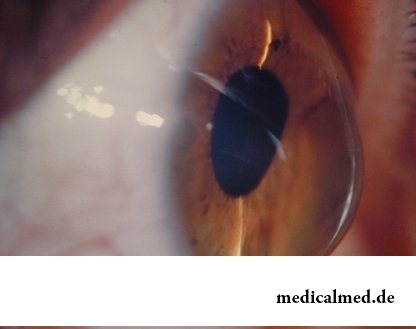
Keratoconus (from Greek "karato" - a cornea and "konus") – such condition of an eye at which the cornea takes conical shape. Under the influence of degenerative processes cells of one of cornea layers (Bouman's layer) collapse that leads to loss of rigidity of a cornea, and it under pressure of intraocular liquid is stuck out outside. The keratoconus leads to strong deterioration in sight, but almost never leads to its full loss.
The keratoconus meets not so seldom, according to medical statistics, 1 person from 2000 gets sick with it. Frequency of a disease does not depend from sexual or race, the first symptoms of a keratoconus are shown, as a rule, at people from the moment of puberty and up to 20 years.
Keratoconus origins
The reasons of development of the degenerative processes in a cornea leading to a keratoconus are still unknown. Influence of autoimmune processes at which cells of immune system destroy own cells of an organism is undoubted. These data are confirmed by the fact that often the keratoconus arises at the people having bronchial asthma, allergies and other frustration of immune system.
One of the factors promoting development of a keratoconus is long reception of corticosteroid drugs that renders, including, influence and on immune system, and also confirms its leading role in developing of a disease.
Dependence of frequency of emergence of a keratoconus on an adverse ecological situation, in particular, long stay in rooms where air is contaminated by a rough suspension of the dust causing constant microtraumas of a cornea is noted. Also there are data on influence of genetic factors on development of a disease. In the majority of cases the reason of emergence of a keratoconus remains obscure.
Keratoconus symptoms
As a rule, keratoconus symptoms appear on one eye in the beginning, but then the second eye is involved in process. The keratoconus seldom happens only on one eye, just different extents of its manifestation are more often on different eyes. The main symptom of a keratoconus is deterioration in sight. In the beginning patients note deterioration in sight in night-time, then blurring of the image is shown also at good lighting. Eyes quickly are tired, sometimes in them unpleasant feelings in the form of an itch or burning appear.
Patients with a keratoconus describe the image before eyes as the vague picture which they see as if through glass during a heavy rain. The image appears doubled, the symptom of a monocular polyopia, characteristic of a keratoconus, appears – when instead of one image the patient sees a little. It is especially expressed when examining light objects against a dark background. To the patient suggest to look at the image of a white point on the black sheet of paper, and instead of one white point in the center he sees several such points which are chaotically scattered according to the sheet of paper and when checking after a while this chaotic sequence does not change.
The patient experiences difficulties at selection of the adjusting points, there is a need for their frequent change.
Usually symptoms of a keratoconus accrue within several months or even years, then the disease ceases to progress and remains at the same level for a long time. In rare instances the keratoconus progresses continuously, leading to frequent ruptures of a cornea and threat of loss of an eye.
Diagnosis of a keratoconus
In the beginning the doctor carefully asks the patient on symptoms, then check visual acuity. Inspection by means of a slit lamp is performed, detection at the same time of a symptom, characteristic of a keratoconus, – "Fleyshner's rings" is one of diagnostic characters. Apply a skiaskopiya – a method of a research at which the ray of light is directed to an iris of the eye of an eye of the patient and, displacing it, monitor reflection. At a keratoconus there is a so-called effect of scissors at which two reflected strips of light move, like edges of scissors.
One of the most informative and exact modern diagnostic methods of a keratoconus is use of the optical topographer, with drawing up the topographic map of a front and back wall of a cornea. Such method allows to diagnose a keratoconus even at early stages, and drawing up topographic maps of a cornea through certain periods allows to observe process in dynamics.
Treatment of a keratoconus
 Depending on features of course of process (bystry progressing, tendency to a recurrence, or on the contrary, slow increase of symptoms with the long periods of stability) there can be both a surgical, and non-invasive treatment of a keratoconus.
From conservative methods of treatment of a keratoconus the following is applied:
- Correction by semifixed lenses is based on mechanical cave-in of a cone of a cornea by means of special lenses, rigid in the center and soft on the periphery;
- Krosslinking (Corneal Collagen Crosslinking, CCL, CXL) – rather new, but perfectly proved method allowing to strengthen a cornea and to increase its rigidity for 300% in the non-invasive way. The essence of a method consists in removal of superficial cells of a cornea, instillation Riboflavinum with the subsequent 30-minute radiation of an eye an ultra-violet lamp. Then the special contact lens protecting a cornea for that time is approximately put on until there are regeneration processes. Such treatment of a keratoconus safely and malotravmatichno, is carried out in out-patient conditions and does not demand use of the general anesthesia.
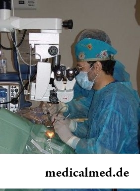
At a heavy current and far come keratoconus stage operation nevertheless is necessary as because of frequent ruptures of a cornea there is a threat of loss of an eye. Today two forms of operations at a keratoconus are taken:
- Implantation in a cornea of intrastromalny corneal ring segments (X), the thin arches from polymer located on both sides from a pupil which put the constant pressure therefore the cone is flattened.
- The keratoplasty, or through keratoplasty – a classical method of operation at a keratoconus when own thinned cornea is removed, and on its place is implanted donor.
Treatment of a keratoconus folk remedies
At treatment of a keratoconus folk remedies are used as fortifying, and also preventive, for avoiding of complications of a disease, such as rupture of a cornea. At an itch, burning and bystry fatigue of eyes recommend washing and lotions broths of medicative herbs, such as a camomile, a sage, coltsfoot. Medicative herbs can be also applied as teas to soft immunocorrection, for example, tea from leaves of a purple cone-flower purple.
One more of folk remedies of treatment of a keratoconus – honey and other products of beekeeping. Honey and propolis in this case is applied as locally, in the form of aqueous solutions, and in food, as fortifying and immunoexcitant.
The educated person is less subject to brain diseases. Intellectual activity promotes formation of the additional fabric compensating sick.

For the time being the perspective of heart diseases seems to most of people remote and foggy. But sooner or later практичес...
Section: Articles about health
During foot walks blood moves on vessels more actively and one and all bodies are supplied with a large amount of oxygen. It affects the state of health of the person very positively....
Section: Slideshow
Coffee – favourite drink of many. For the last decades it more than once already declared very harmful, extremely useful and even necessary for normal life activity. In spite of the fact that this product became for us usual for a long time, there are many myths about properties of coffee and its impact on a human body. Readers can get acquainted with the most widespread of similar delusions today....
Section: Articles about health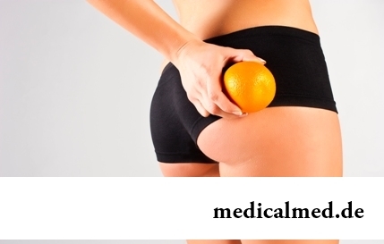
Cellulitis - very widespread cosmetic shortcoming which arises approximately at 80% of women sooner or later. Emergence ег...
Section: Articles about health
Shops of household appliances offer us the huge choice of various devices for the house. Whether there are among this abundance devices which not only facilitate house work, but also help to keep health of the person? Of course, and we will tell about them today....
Section: Articles about health
For the person who daily since morning gathers for work it is very important to wake up vigorous and ready by day of work. Actually, each of us experiences difficulties with this, at first sight, simple business from time to time. After night rest exert impact on a condition of an organism the weather which collected for several days fatigue, household and office problems, quality of a dream and many other factors....
Section: Articles about health
What will only not be thought up by persons interested to have a beautiful figure. Here the last innovation – for weight loss needs to be eaten greasy food. Give ра...
Section: Slideshow
Color of plants is caused by presence at them of certain chemical compounds. Let's talk about what is meant by various colors of vegetables and fruit and what properties they give them....
Section: Articles about health
Partial and the more so full loss of hearing significantly reduces quality of life. Difficulties with communication lead to loneliness and isolation. The person who badly hears experiences difficulties with social and professional implementation, quite often has problems in private life....
Section: Articles about health
Heart disease and blood vessels lead to disturbance of blood supply of bodies and fabrics that involves failures in their works...
Section: Articles about health
When overcomes feeling of hunger, and an opportunity to have dinner fully is absent, having a snack − the meals, small on volume, stabilizing sugar level in blood comes to the rescue. The relation of nutritionists to having a snack is more often negative, but only because in кач...
Section: Articles about health
Condition of lips (their morbidity, outward) – one of indicators of health of the person. The peeling, dryness, pallor, and also cracks in corners of a mouth can be not only the cosmetic shortcoming which arose owing to physical damages and weather conditions but also the satellite of some diseases and disturbances in an organism needing treatment. Let's consider 10 possible reasons of emergence of angular cracks (perleches) in corners of a mouth and ways of their elimination....
Section: Articles about health
Impossibility to conceive the child – a trouble of many Russian families. During quite long time was considered that the main "culprits...
Section: Articles about health
Separate food - the system of meal based on digestion physiology which is carried to improvement methods. According to nutritionists, the separate use of the carbohydrate and proteinaceous products demanding different conditions of assimilation helps to get rid from Bol...
Section: Articles about health
The summer of this year in Russia was very ambiguous. Regions suffered from a merciless heat, from pouring rains, the hail from time to time dropped out, then there was again a heat which alternated with rainfall again. Many people suffer from such sharp changes of weather. Even flu epidemics and a SARS were recorded....
Section: Articles about health
The state of health of the person in many respects depends on chemical composition of biological liquids of an organism. Specialists consider that з...
Section: Articles about health
Reactive pancreatitis - the disease which is characterized by inflammatory process in a pancreas which arises most often because of excess activity of digestive enzymes. It − the emergency state which treatment has to take place in хирургич...
Section: Articles about health
Eyes – unique body on the structure thanks to which the person obtains about 80% of information on the world around: about a form, color, size, the movement, and also many other parameters of objects or phenomena. But whether much we know about the most valuable sense body which, according to the scientist Sechenov, provides us about one thousand various feelings a minute? Let's consider 10 most surprising facts about eyes and sight....
Section: Articles about health
Cold – a state known to everyone which is followed by cold, cough, high temperature, a pharyngalgia. Often перво...
Section: Articles about health
It seems, quite recently you brought the baby from maternity hospital, but time flew by, and here it is already going to join the first in life children's collective. How to prepare the child for visit of a garden? What needs to teach him to facilitate process адап...
Section: Articles about health
It would seem, to buy drugs in Moscow does not make a problem – a drugstore, and not one, is available for each resident of the capital within walking distance. And, nevertheless, Internet drugstores become more popular – what it is possible to explain such phenomenon with? Actually there is a lot of reasons and if to formulate them it is short, then the most suitable word will be - "conveniently". We suggest to get acquainted in more detail with pluses and minuses of online drugstores that buying drugs, not to make the wrong choice....
Section: Articles about health
The chia plant, or the Spanish sage, is from South America. The indigenous people of the continent since ancient times used in food it семена:...
Section: Articles about health
What woman does not dream of a beautiful and thick hair? While physicians developed difficult schemes on hair transplant, in the industry of hairdresser's art a few years ago there was a sensation – methods of hair extension appeared. It would seem, dreams came true...
Section: Articles about health
Olive oil – the product capable to make a powerful contribution to health of the person if it includes it in the diet. The rich vitamin composition of oil does it by a product number one from many diseases including from deadly. Only two tablespoons of oil from olives in day prevent emergence of diseases of vessels and heart, cancer, problems with digestion, presenilation, a depression and many other illnesses which treatment would demand a lot of time and forces. Let's consider on...
Section: Articles about health
Wood louse – the ordinary-looking unpretentious plant extended in all territory of our country. It quickly expands, and sometimes for...
Section: Articles about health
The list of stereotypes of which, apparently, all know strongly includes following: British surely eat porridge for breakfast. Perhaps, not all modern residents of Britain arrive quite so, but for those from them which continue to follow this t...
Section: Articles about health
About 10-15 years ago existence of the computer in the apartment of the Russian was considered as a rarity and office rooms were only at the first stage of equipment by these useful devices. Today practically in each house there is a computer (and often not one), and a regular user is already every our second compatriot. Convenience and efficiency of personal computers are undoubted, but the people working with them daily have to know also about health hazard which they can predstavlit...
Section: Articles about health