Deratofibroma
The deratofibroma represents high-quality education on skin of the small size. The deratofibroma can appear on any body part, but shoulders, an upper back, a foot and an anklebone are the most widespread. It is possible to meet education at adults more often, at children – is much more rare. Education can be gray, pink, brown or red color, sometimes over time it changes color. To the touch the deratofibroma firm also reminds the lump which hardened under skin. The skin outgrowth is preferential formed by fibrous fibrous fabric and contains histiocytes and fibroblasts. If to squeeze a deratofibroma on the parties, then on its surface the small pole will appear. The deratofibroma is called also sclerosing hemangioma or a fibrous histiocytoma.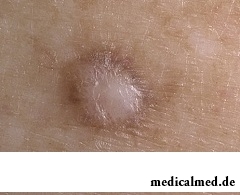
As a rule, deratofibromas do not cause essential inconveniences. They are almost painless, but can cause an itch and weak pain in some people. It is most often possible to see single deratofibromas, but at some patients it is possible to meet also multiple. Educations seldom happen more than one centimeter in the diameter.
Deratofibroma reasons
Was considered earlier that the deratofibroma is reaction to stings of various insects or an injury, however this version did not gain distribution. The exact reason of a deratofibroma is unknown. Partly it can be explained with the resistant nature of a disease. At first education reminds a dense small knot, the size in a small pea. Eventually the deratofibroma size, its color and density change a little.
Doctors allocate such probable causes of a deratofibroma:
- Sexual predisposition. Women are more subject to a disease, than men.
- Injury of skin. The deratofibroma can result from sting, a thorn prick, etc.
- Hereditary factor. A cause of illness it becomes frequent a hereditary family factor.
- Age. Most often the deratofibroma is formed at middle-aged persons. Occurs at children of a disease seldom.
Deratofibroma symptoms
The most common symptoms of a deratofibroma are:
- the consolidation arising over the surface of skin which at damage can bleed;
- new growths are most often formed standing in the area below a knee, but can appear on hands and a trunk;
- when pressing the deratofibroma caves in inside;
- color of a deratofibroma significantly differs from a normal shade of skin;
- at people with swarty skin more dark shade, as a rule, has education; at a touch to a new growth there can
- sometimes be a morbidity and an itch.
It is necessary to know that very often the dermatofibrosarcoma (a kind of a carcinoma cutaneum) at the first stage has similar symptoms with a deratofibroma. Therefore it is important not to play for time, and at manifestation of the first symptoms to see a doctor. Especially it is worth paying attention when education quickly increases in a size, changes color or a form, causes strong pain or bleeds.
Diagnosis of a deratofibroma
The doctor, as a rule, can diagnose a deratofibroma after visual survey of the patient. If the outgrowth on skin does not bleed and visually is not similar to a deratofibroma, the doctor can make a biopsy. For this purpose the doctor will remove a small piece of education and will carefully study it under a microscope. By means of a biopsy it is possible to diagnose various diseases including tumors. Before carrying out a biopsy the patient is exposed to a local anesthesia.
Treatment of a deratofibroma
 The disease in itself does not pass, and without the corresponding treatment the deratofibroma remains with the patient throughout all his life. If educations reside in too noticeable places or the patient touches them (in the course of clothing or at commission of other actions), then removal of a deratofibroma is more preferable. It is also best of all to choose removal of a deratofibroma if it causes at the patient unpleasant feelings, morbidity and an itch.
As deratofibromas are deeply under skin, for their removal do cuts in deep layers of skin. After removal of deratofibromas often there are quite noticeable scars. If the procedure takes place normally, the patient is allowed to go home in the same day. Complications develop extremely seldom and they are connected, as a rule, not with a disease, and with poor carried out procedure.
Alternative option of treatment of a deratofibroma is cutting of its upper part. But most often after such treatment it increases in a size several years again later.
After removal of a deratofibroma the scar for some time remains very noticeable, often causes an itch. Over time it smoothes out and becomes more pale. Today there is a set of cosmetic procedures which allow to make a scar practical imperceptible. However it is necessary to know that it is possible to undergo these procedures only after a while after treatment of a deratofibroma. For treatment of hems it is possible to choose one of the following methods:
- laser treatment by means of which it is possible to eliminate reddening effectively;
- injections of steroids in a hem;
- the special silicone gel helping to smooth edges of a hem;
- at very big hems it is possible to resort to surgery.
During life the average person develops neither more nor less two big pools of saliva.

The phenomenon of improvement of a condition of the patients at administration of drugs who are not containing active agents, so-called effect of placebo is known...
Section: Articles about health
When overcomes feeling of hunger, and an opportunity to have dinner fully is absent, having a snack − the meals, small on volume, stabilizing sugar level in blood comes to the rescue. The relation of nutritionists to having a snack is more often negative, but only because in кач...
Section: Articles about health
Partial and the more so full loss of hearing significantly reduces quality of life. Difficulties with communication lead to loneliness and isolation. The person who badly hears experiences difficulties with social and professional implementation, quite often has problems in private life....
Section: Articles about health
Weakness of an ankle joint – very widespread problem. Its existence is demonstrated by tendency to a podvorachivaniye of legs п...
Section: Articles about health
Antibiotics - - it is possible to call the chemical compounds suppressing growth of bacteria the break in the field of medicine which allowed to save mankind from many diseases incurable earlier: tuberculosis, plague, syphilis and many others. A contribution of drugs to rescue of people from...
Section: Articles about health
According to World Health Organization, every third inhabitant of Earth has excess weight, and every tenth has obesity. The reason of this phenomenon, according to specialists, roots in one not very comforting fact: most of people consume much more calories, than it is necessary. How it turns out what we overeat? Why it is so difficult to refuse an excess portion tasty or additives? Let's try to find out what factors prevent us to eat food with reasonable moderation....
Section: Articles about health
Use of medicinal plants in therapy is urgent today, more than ever. The drugs made of curative herbs cannot on...
Section: Articles about health
The endocrine system carries out extremely important role in a human body, practically all processes of life activity are regulated by it. Closed glands (hemadens) produce special biologically active agents – hormones which then o...
Section: Articles about health
What they, women? Beautiful, gentle, passionate and at the same time windy, gusty, and nervous. And what is stranger: have all these qualities of the woman at the same time. But here only the mood their time sharply changes on completely opposite: in the morning they laugh and joke, and in the evening cry or are irritated....
Section: Articles about health
About 10-15 years ago existence of the computer in the apartment of the Russian was considered as a rarity and office rooms were only on перв...
Section: Articles about health
Stability of a hormonal background is one of the most important conditions of preservation of health of the woman. At the same time endocrine system – the thin device extremely sensitive to any external influences. Changes of an image жиз can become the reason of hormonal failure...
Section: Articles about health
Practically each person is familiar with the annoying, pulling, unscrewing pains caused by overcooling of muscles of a back. In certain cases inflammatory process is not limited to discomfort, being followed by emergence of hypostasis, consolidations, temperature increase. At the wrong treatment the acute miositis can lead to a chronic disease or aggravation of other pathologies of a back (vertebral hernia, osteochondrosis) therefore it is important to pay attention to symptoms of an illness in time and to start to...
Section: Articles about health
Statistically, at the address to doctors seven of each ten patients complain of a headache. Actually people, periodically...
Section: Articles about health
The pancreas performs two functions in a human body: release of enzymes without which digestion of carbohydrates, fats and proteins, and a producing hormones is impossible. The most important of them - insulin, is the main participant of carbohydrate metabolism, a normal...
Section: Articles about health
A little more than a century ago goat milk was a traditional food stuff of most of Russians. Unfortunately, today on tables of our compatriots it appears extremely seldom. The reason that the use of so useful product practically came to naught, not only in very modest volumes of its production and, respectively, rather high cost. Potential consumers are just insufficiently informed on unique properties of goat milk and that advantage which...
Section: Articles about health
Cold – a state known to everyone which is followed by cold, cough, high temperature, a pharyngalgia. Often перво...
Section: Articles about health
History of cultivation of a buckwheat contains more than five thousand years. Grain which is received from this plant is used for preparation of porridges, soups, baked puddings and puddings, do flour which is one of the main ingredients of the noodles popular in of it...
Section: Articles about health
The words "disease" and "patient" not without reason come from one root – "pain". As a rule, symptoms of illnesses thoroughly spoil to patients life. However from this rule there are exceptions. Some diseases are shown by signs which can cause even positive emotions. It is a pity only that the majority of such illnesses are heavy and incurable....
Section: Articles about health
The state of health of the person in many respects depends on chemical composition of biological liquids of an organism. Specialists consider that з...
Section: Articles about health
So, you resolved to lose weight. And now you try to understand what to begin with: from exercise stresses or a diet? And how to make that process of weight loss did not give you an inconvenience, and, on the contrary, brought joy?...
Section: Slideshow
It seems, quite recently you brought the baby from maternity hospital, but time flew by, and here it is already going to join the first in life children's collective. How to prepare the child for visit of a garden? What needs to teach him to facilitate adaptation process? What to tell and how to behave that the kid transferred changes in the life without serious consequences? Let's try to find answers to these questions....
Section: Articles about health
Aging — natural and inevitable process. Over time our skin loses elasticity, on it saggings, a face form теря are formed...
Section: Articles about health
On health of the person physicians know about salutary action of animals long ago. About 7 thousand years ago great Hippocrates recommended to the patients riding walks for strengthening of a nervous system and increase in vitality....
Section: Articles about health
Very often as a source of the infection which caused a disease serves our house - the place which a priori has to be safe. However disease-producing bacteria can perfectly feel not only in insanitary conditions, but also in our apartment if not to carry out due care of favourite places of their dwelling. What they − sources of their reproduction? Let's consider 10 most widespread places in our house, the most dangerous from the point of view of infection with microorganisms....
Section: Articles about health
Diapers for adults – individual one-time means of hygiene which in some situations is irreplaceable, and from such situats...
Section: Articles about health
Dogrose – one of the most widespread adornment and medicinal plants growing practically in all territory of our country. To most of Russians it is a beautiful bush it is known, first of all, as a source of fruits, extremely vitamin-rich....
Section: Articles about health
Life of the modern child is extremely active and difficult. Information strain which is experienced by the school student and did not dream pupils of last times. Careful parents, wishing well to the children, will organize a set of additional classes in circles, sports sections and music schools. In such situation the child needs continuous care and good nutrition to keep health and high performance....
Section: Articles about health