Humeral bone
Humeral bone – a skeletal basis of a shoulder, a long tubular bone.
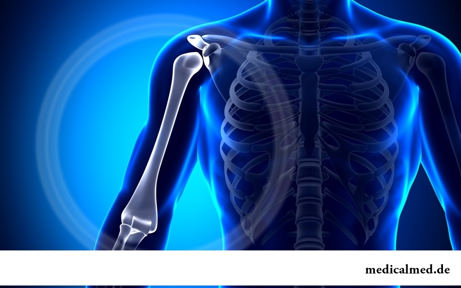
Structure of a humeral bone
The humeral bone consists of a body and two epiphysis – distal lower and proximal upper.
In a lower part of a body of a bone there is a back surface limited on the periphery medial and lateral to edges, and also the lateral and medial front surfaces divided by slightly noticeable crest.
On a medial front body surface, is slightly lower than a middle part of a body, there is a nutritious opening conducting in distally the directed nutrient canal.
On a lateral front surface, is slightly higher than a nutritious opening, it is possible to see deltoid tuberosity – the place where the deltoid muscle fastens.
Behind deltoid tuberosity, on a back body surface, there passes the furrow of a beam nerve.
The proximal epiphysis is a little thickened. On it there is a semi-spherical head of a humeral bone turned up, inside and it is a little back. From other part of a bone the periphery of a head is limited to the small narrowing going annularly, a so-called anatomic neck. A little below it two hillocks – small and big are located. Down from each hillock last respectively a crest small and a crest of a big hillock. They are directed down and reach upper parts of a body of a bone. Together with hillocks they limit accurately expressed intergrumous furrow in which there is a sinew of a long head of a two-headed humeral muscle.
On border of a body and the upper end of a bone, is a little lower than hillocks, there is a surgical neck – the small narrowing corresponding to an epiphysis zone.
The distal epiphysis is squeezed in the perednezadny direction. Its lower part is called a condyle of a humeral bone. The condyle consists of a head to which the head of a beam bone, and the block which connects in an elbow joint to blokovidny cutting of an ulna connects.
In front of a distal epiphysis it is possible to see a coronal pole, over a condyle head – a beam pole, and on a back surface – a pole of an elbow shoot.
Peripheral departments of the lower part of a bone come to an end medial and lateral with epicondyles from which forearm muscles originate.
From each epicondyle lateral and medial epicondylic crests climb distal department respectively.
The medial epicondyle is stronger developed. On its dorsum it is possible to see a furrow of an elbow nerve, and in front there is a ledge – an epicondylic shoot with which the beam sgibatel of a wrist begins.
The furrow of an elbow nerve and epicondyles are well probed under skin and serve as bone reference points.
Fractures of a humeral bone
There are following types of fractures of humeral bone:
- Head change;
- Intra articulate change (fracture of an anatomic neck);
- Extraarticular changes (chrezbugorkovy fracture and fracture of a surgical neck);
- Change of a hillock of a humeral bone.
Changes of a head and anatomic neck of a bone arise, as a rule, as a result of direct stroke to the outside surface of a shoulder joint, or as a result of falling on an elbow joint. At the same time the head of a bone breaks up into several parts.
The clinical picture of a change is characterized by emergence of sharp pain. At the expense of hypostasis the shoulder joint increases, there is no opportunity to make the active movements by a hand. The passive movements are painful.
Fractures of a surgical neck of a humeral bone divide on abduction (taking-away) and adduktsionny (bringing).
The Adduktsionny fracture of a neck of a humeral bone arises preferential when falling with the emphasis on the extended given hand, and abduction – when falling with the emphasis on the extended taken-away hand.
At a fracture of a neck of a humeral bone without shift the patient feels the localized pain which at axial loading amplifies. At the same time function of a shoulder joint is limited.
At a change with shift the patient feels sharp pain and pathological mobility. Function of a shoulder joint is broken, the axis of a shoulder is broken and shortened.
The change of a hillock of a humeral bone most often occurs at dislocation of a shoulder or at the indirect mechanism of an injury. The change results from reflex reduction small round, podostny and supraspinal muscles. The isolated change of a hillock of a humeral bone without shift, as a rule, results from a shoulder bruise.
At a change patients feel the localized pain, there is hypostasis of soft tissues. The active movements cannot be carried out.
In Great Britain there is a law according to which the surgeon can refuse to do to the patient operation if he smokes or has excess weight. The person has to refuse addictions, and then, perhaps, he will not need an operative measure.

The pancreas performs two functions in a human body: release of enzymes without which digestion carbohydrate is impossible...
Section: Articles about health
It is pleasant to state a possibility of improvement of quality of life of people with problems of functioning of secretory system. Efforts of talented inventors created products which will be able to provide normal life activity of clients with moderate degree for...
Section: Articles about health
Subfebrile temperature call fervescence to 38 degrees, and subfebrile condition - existence of such temperature over 3 days, and quite often it happens without the visible reasons. Existence of subfebrile condition - a strong indication of disturbances in an organism which can be caused by various reasons: disease, stresses, hormonal failures. Despite the seeming inoffensiveness it is a state at which people often continue to lead a usual life, often is a sign of many of a zabolev...
Section: Articles about health
Helminthosis is one of the most widespread diseases. Statistically, with any species of helminths it is infected porridges...
Section: Articles about health
For anybody not a secret that the modern person eats not as his ancestors. For the last 100 years in broad access there were absolutely new products which are result of use of the latest technologies in food production. Significantly changed спо...
Section: Articles about health
White teeth and the Hollywood smile – a dream of many people. Long time was considered that the plaque on teeth and change of their color – destiny of those who incorrectly eat smokes and badly brushes teeth. But the paradox is available: at everything the variety of toothpastes, brushes existing today for toothbrushing and conditioners for a mouth the number of the people hesitating of a plaque on teeth does not decrease. Moreover, the plaque is formed even at small children who definitely do not smoke and have no coffee. So in what business, and опас...
Section: Articles about health
When overcomes feeling of hunger, and an opportunity to have dinner fully is absent, having a snack − small on volume comes to the rescue...
Section: Articles about health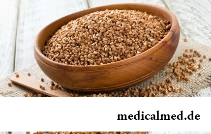
History of cultivation of a buckwheat contains more than five thousand years. Grain which is received from this plant is used for preparation of porridges, soups, baked puddings and puddings, do flour which is one of the main ingredients of the noodles popular in of it...
Section: Articles about health
About influence of fasting days on an organism it is told much – both about advantages, and about shortcomings. It is considered that fasting day in the form of a short-term monodiet is useful, promoting effective removal of slags from an organism whereas irregular, excessively long, spontaneous fasting days lead only to deterioration in health. How to derive benefit from the sparing diet and not to do much harm to itself? Let's consider the main advantages and shortcomings of fasting days and their influence on an org...
Section: Articles about health
Water with a lemon - idle time in preparation drink which supporters of a healthy lifestyle already managed to appreciate. Upo...
Section: Articles about health
The person, as well as all other beings living on our planet feels weather changing. It is the normal meteosensitivity which is not causing to healthy people of special troubles. Meteodependence, on the contrary, is morbid condition, характеризующимс...
Section: Articles about health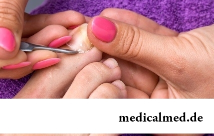
Beauty shop – the place which is associated only with positive emotions: joy, pleasure, relaxation. However visit of salon where work with biological material of clients, not always harmlessly is conducted. Today more than 100 pathogenic microorganisms who can catch in beauty shop including deadly to health are known....
Section: Articles about health
All diseases from nerves – in this joke a big element of truth, are said by doctors. Constant stresses lead body to decrease in protective forces...
Section: Articles about health
The concept "gluten" (differently, a gluten) combines group of the proteins which are a part of rye, barley and wheat. For most of people the use of the food stuffs containing a gluten not only is safe, but also it is very useful. Nevertheless, there is a number the myth...
Section: Articles about health
The sudden heat on all body which is followed by perspiration and a cardiopalmus – the phenomenon familiar to many people. Most often such states called by "inflows" result from nervous or physical overworks and disappear right after rest. However in certain cases similar reaction of an organism can speak about diseases which need treatment. What? About it below....
Section: Articles about health
For residents of the countries of Southeast Asia various algas are an obligatory component of a daily diet. Their priest...
Section: Articles about health
Long time antibiotics were considered as a panacea from all diseases and were appointed even at insignificant symptoms of an infection. Even now not everyone knows in what force of antibiotics how and when they should be accepted. Let's discredit 7 popular myths about such drugs...
Section: Articles about health
Life of the modern woman is very difficult. Opportunities to realize itself are wide: it not only education and career, but also the most various hobbies from sport before needlework. It is not less important to build private life, paying an attention maximum to children, the husband, parents, friends. For all these affairs there is catastrophically not enough time therefore each of us tries to cut down as far as possible its expenses on necessary, but not the most fascinating housework. With it we are successfully helped by means...
Section: Articles about health
Many of us, probably, noticed more than once that from intellectual loadings at some point the brain as though "overheats" also "assimilation"...
Section: Articles about health
Neurosis is called pathology of a nervous system at which deviations in functioning of the highest nervous processes are observed. Most often - owing to yet not strengthened mentality - children are subject to neurosises. Premises to emergence of such disturbances can become нез...
Section: Articles about health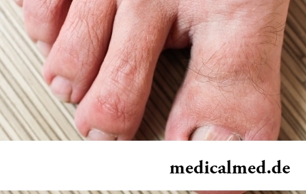
The word "onikhokriptoz" is unfamiliar to most of people, meanwhile quite so physicians call very widespread problem: the growing of edge of a nail into surrounding fabrics causing inflammatory process. Usually the illness affects thumbs of legs, and is followed by reddening, hypostasis, and in the started cases – release of pus. Patients complain of the pain amplifying when walking, problems with the choice of footwear....
Section: Articles about health
Aspirin (acetylsalicylic acid) – one of those drugs which are known literally to all. It is available in each home first-aid kit...
Section: Articles about health
The advantage of swimming for the person is so high that this sport is not only the most popular, but also is widely applied in medicine and rehabilitation processes. If you look for for yourself the occupation allowing pleasantly and to spend time, then swimming with advantage...
Section: Slideshow
Statistically, pathologies of a thyroid gland in the world more than 500 million people have. Failures in work of this body lead to heavy disbolism, development of heart diseases, vessels, a reproductive and nervous system. In hard cases excess or insufficient production of the main hormones of a thyroid gland (thyroxine and triiodothyronine) leads to essential decline in quality of life and disability....
Section: Articles about health
For most of the working people the problem of having a snack is particularly acute enough. Sooner or later there is a question: what is possible quickly for a sja...
Section: Articles about health
Several decades ago the basil (the district khan, реан, Reagan) was considered as a part of the Caucasian or east cuisine, but today it strongly took the place on tables of Russians. Greens of this plant possess a strong, pleasant smell and specific fresh taste, because of to...
Section: Articles about health
Quite large number of people adheres to the principles of vegetarian food. But how to be if in a family of vegetarians there are children? Whether it is possible to eat also it the same as to parents, or after all the children's organism is not adapted for the use of exclusively vegetable food? Let's try to understand....
Section: Articles about health