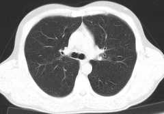
Tomography of lungs
 Tomography of lungs - one of ways of diagnosis of diseases of lungs. Carry out it by means of the ring-shaped X-ray doing pictures of lungs under different corners. Big distribution was gained by a computer tomography which gives the chance to digitize the received pictures, to provide numerous cuts of lungs for the analysis.
It is possible to make a tomography of lungs in two modes: to estimate a condition of lungs and bodies of a mediastinum: tracheas, hearts, upper vena cava, lymph nodes, pulmonary aorta and artery.
In some cases, when there are difficulties with diagnosis, to the patient enter contrast better to consider lungs.
By means of a tomography it is possible to reveal a chronic embolism, tuberculosis, diffusion diseases, lung cancer (localization of tumors, a condition of lymph nodes, existence and prevalence a metastasis), the pneumonia, occupational diseases caused by inhalation of silicon particles, asbestos, quartz, etc.
Indications to purpose of a tomography
To make a tomography of lungs appoint in such states and suspicion to them:
- change of structure of tissue of lungs;
- disturbances in work of a thymus,
- liquid in a pleura;
- inflammation of lymph chest nodes;
- diseases of a breast and edges;
- changes of a pericardium;
- new growths of the high-quality and malignant nature in lungs and a pleura
- foreign bodys in lungs;
- bronchoectatic disease
Preparation for a tomography of lungs
At purpose of such inspection patients need to warn the doctor about pregnancy, feeding by a breast, heart diseases, asthma, problems with a thyroid gland, about existence of a myeloma in the anamnesis, claustrophobia.
 The tomography of lungs – the procedure painless and short, but to the patient at the increased emotional excitability and concern caused by the outlined inspection is allowed to accept soothing.
In certain cases practice anesthesia before a tomography to provide the patient's immovability: children and "heavy" patients.
Just before the procedure the patient has to undress to a belt, take off from himself the jewelry and objects containing metal, to lay down on a table. It is impossible to move to inspection time. Existence of pacemakers is not a contraindication to carrying out a tomography.
The patient is in a radiological office one, and the technologist who watches the equipment is in the next office. Communication between them happens by means of an intercom.
Results of a tomography are ready right after the end of inspection and the radiologist can read the main diagnosis to the patient. The detailed conclusion in writing can be taken away only in 1-2 days.
It is possible to carry out a tomography easy for children?
Considering that the tomography assumes radiation of the patient, at purpose of a tomography easy for children, not all parents consider it expedient and are afraid of serious effects of influence of radiological beams.
Appointing a tomography easy for children, the doctor recognizes those reasons that the dose of radiation is small, the procedure lasts only 1-7 minutes, and cannot constitute danger, and the advantage of timely diagnosis many times over exceeds presumable harm from radiation. Therefore it is even authorized to newborn children to appoint such inspection.
The magnetic and resonant tomography - the research based on ability of radiation by hydrogen of electromagnetic waves can become an alternative of a tomography of lungs and to radiation. The possibility of use of this method depends on specifics of a disease and it should be discussed with the attending physician.
People who got used to have breakfast regularly have obesity much less often.

The trophic ulcer is not an independent disease. This heavy complication arising owing to a thermal injury (a burn...
Section: Articles about health
Bees – really unique beings. Practically all products of their life activity are used by the person. Since the most ancient times medicinal properties of honey and other substances received in the course of beekeeping are known. The fact that all these пр is especially significant...
Section: Articles about health
Color of plants is caused by presence at them of certain chemical compounds. Let's talk about what is meant by various colors of vegetables and fruit and what properties they give them....
Section: Articles about health
Modern footwear is extremely various. It stopped being only protection for legs long ago. Today shoes, boots, barefoot persons in...
Section: Articles about health
The pine is one of the most widespread plants of our woods. Its needles and pitch not without reason called by "gallipot" were since ancient times used for strengthening of protective forces of an organism, treatment of avitaminosis, anemia and many other diseases. In recent years wide п...
Section: Articles about health
The cosmetics intended for improvement of a condition of skin, nails and hair are used by each woman. Expenses on regular acquisition of the fashionable widely advertized products of well-known companies for many become very notable and significantly burden the family budget. Meanwhile, there is a number of inexpensive pharmaceutical drugs which can quite be applied in the cosmetic purposes. At the same time the effect of their use is often more noticeable, than result of use of the most expensive...
Section: Articles about health
The name of this disease precisely reflects the problem reason: it consists in the bra fastener pressure upon a certain zone...
Section: Articles about health
One of the major chemical processes happening in a human body are oxidation reactions. They go with participation of fats and carbohydrates which we receive from food, and the oxygen getting to us from air. A main goal of such reactions is it is received...
Section: Articles about health
Each person has easy indispositions which he transfers "standing", trying not to ask for medical care. Arguments at the same time are adduced same: "it is a trifle, itself will pass", "I have too many important issues", "there are no wish to spend time for doctors", etc. At good shape of health, normal working capacity and lack of suspiciousness dislike for complaints to such problems is quite natural. It is not the most correct, but very widespread type of behavior. I am glad...
Section: Articles about health
Impossibility to conceive the child – a trouble of many Russian families. During quite long time was considered that the main "culprits...
Section: Articles about health
Smack in a mouth can arise in the natural way – as a result of lack of morning hygiene or reception of the corresponding food. However in certain cases its existence is a sign of certain pathologies, and allows to reveal an illness at an early stage. In we depend...
Section: Articles about health
It would seem, to buy drugs in Moscow does not make a problem – a drugstore, and not one, is available for each resident of the capital within walking distance. And, nevertheless, Internet drugstores become more popular – what it is possible to explain such phenomenon with? Actually there is a lot of reasons and if to formulate them it is short, then the most suitable word will be - "conveniently". We suggest to get acquainted in more detail with pluses and minuses of online drugstores that buying drugs, not to make the wrong choice....
Section: Articles about health
For many spouses the question of planning of a family is one of the main. The choice problem effect at the same time comes out on top...
Section: Articles about health
Reactive pancreatitis - the disease which is characterized by inflammatory process in a pancreas which arises most often because of excess activity of digestive enzymes. It − the emergency state which treatment has to take place in хирургич...
Section: Articles about health
Statistically, in Russia about 34% of citizens smoke. Most of consumers of tobacco has problems with health sooner or later. Not only smokers, but also their relatives suffer. Besides, cigarettes are expensive, and need of their acquisition goes a heavy burden on the budget of thousands of Russian families. Many people dream to refuse harmful tendency, but everyone manages to make it not: nicotine addiction is affectionate and to get rid of it not easy....
Section: Articles about health
All diseases from nerves – in this joke a big element of truth, are said by doctors. Constant stresses lead body to decrease in protective forces...
Section: Articles about health
Let's begin with the fact that a separate illness which is called "adjournment of salts", just does not exist. In practice this household name of disbolism leading to development of a number of diseases. Pathological process consists that in an organism проис...
Section: Articles about health
For most of the working people the problem of having a snack is particularly acute enough. Sooner or later there is a question: what can be eaten quickly between a breakfast and a lunch or a lunch and leaving from service so that to receive necessary power feed, but not to overload an organism with harmful components or excess calories? We bring to your attention the list of products which quite conform to these requirements....
Section: Articles about health
The dietology, as well as other sciences, does not stand still. Food stuffs are exposed to comprehensive study, and scientists receive new and...
Section: Articles about health
There is a lot of fans of beer in our country. Statistically, on each average Russian (including women and children) in a year about 60 liters of this drink are consumed. It is not a lot of, as in the Czech Republic or Germany, but figure all the same impressive. Radova...
Section: Articles about health
In consciousness of our many compatriots idea that folk remedies if are no more effective, than medicinal "chemistry" strongly took roots, then are precisely less harmful. Unfortunately, it is not always fair: some methods of treatment consecrated with "century national experience" can work so on the patient that it will need urgent intervention of physicians....
Section: Articles about health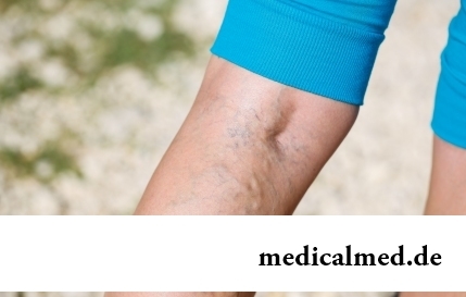
The varicosity has familiarly many, statistically, this disease more than a half of all adult population. As...
Section: Articles about health
Practice of use of table salt in the therapeutic purposes contains not one century. Applications which do by means of the fabric impregnated with saline solution are considered especially effective. They possess antibacterial and antiinflammatory эффек...
Section: Articles about health
The state of health of the person in many respects depends on food. The organism will well function if during food it receive only useful substances, necessary vitamins and microelements. In this case there will be no problems with digestion, with excess weight, and intellectual and physical activity will remain at the high level....
Section: Articles about health
Musicotherapy – a treatment method which caused and causes a set of a controversy concerning its efficiency. However the facts are relentless:...
Section: Articles about health
The words "disease" and "patient" not without reason come from one root – "pain". As a rule, symptoms of illnesses thoroughly spoil to patients life. However from this rule there are exceptions. Some diseases are shown by signs which can cause even полож...
Section: Articles about health