Transvaginal ultrasonography
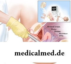 Transvaginal ultrasonography – a method of ultrasonic diagnosis at which the research of bodies of a small pelvis is conducted by the special vulval sensor. Such research is conducted at gynecologic and urological diseases, and also on early durations of gestation. Transvaginal ultrasonography allows to diagnose gynecologic and urological diseases, to diagnose pregnancy on early terms. This type of survey is more informative, than survey through an abdominal wall as in this case the sensor of the device is separated from actually bodies of a small pelvis only by a thin wall of a vagina. Transvaginal ultrasonography of bodies of a small pelvis is eurysynusic, safely, informatively and can repeatedly be carried out.
Indications to transvaginal ultrasonography
Indications to this method of inspection are a suspicion of diseases of bodies of a small pelvis, medical emergencies (for example, an extrauterine pregnancy), control of the carried-out treatment. Transvaginal ultrasonography is carried out at the following states:
- Diagnosis of pregnancy on early terms;
- Pains in lower parts of a stomach;
- Diagnosis of the reasons of infertility and observation of the follicular device of ovaries;
- Disturbances of a menstrual cycle (a delay of periods, bleeding in the middle of a cycle), pathological allocations from a genital tract;
- Detection of inflammatory gynecologic diseases;
- Diagnosis of new growths of a small pelvis, including hysteromyoma, endometriosis, cysts of ovaries, etc.;
- Reception of hormonal drugs, existence of intrauterine contraceptives (spiral) for control of a condition of an endometria and prevention of complications;
- Early durations of gestation, when traditional transabdominal access (through an abdominal wall) малоинформативен;
- Support of the EKO procedure (extracorporal fertilization);
- Installation of the reasons of urological diseases, frustration of an urination, incontience of urine and at pathology of an urethra (urethra).
Transvaginal ultrasonography of bodies of a small pelvis is ideal option at women with obesity as an ordinary research through an abdominal wall at them maloinformativno.
Contraindications
Absolute contraindications to transvaginal ultrasonography do not exist. At virgins survey through a rectum is possible (transrektalno). Transvaginal ultrasonography at pregnancy is justified only on early terms (up to 11-12 weeks).
Preparation for transvaginal ultrasonography
For transvaginal ultrasonography of a uterus and appendages special preparation (filling of a bladder) is not required. At visit of an office of ultrasonography you will need a towel or a diaper on which you will lay down when carrying out a research.
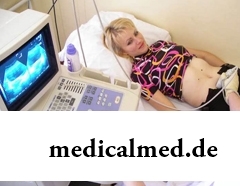 If tranvaginalny ultrasonography at pregnancy is carried out, then the bladder of the patient has to be moderately filled (to drink about 500 ml of liquid for an hour prior to a research).
Indispensable condition for ultrasonography of bodies of a small pelvis transvaginalno is lack of gas in intestines. For this purpose in 2-3 days prior to a research it is necessary to limit the products causing the increased gas generation (vegetables, fruit, bread, fermented milk products, confectionery) and also reception of some medicines reducing gas generation in intestines is recommended – enzistat, absorbent carbon. It is not recommended to carry out cleansing enemas before a research. It is not obligatory to carry out transvaginal ultrasonography of a uterus and appendages on an empty stomach.
In case of emergency transvaginal ultrasonography can be executed also without preparation, however its informational content can be reduced.
Ultrasonic examination of gynecologic bodies is recommended to be conducted in the first half of a menstrual cycle (usually for 5-7 day) as in the second half its endometria of a uterus is in a secretory phase that can result in the wrong interpretation of results. However at endometriosis performance of transvaginal ultrasonography of a uterus in the second phase of a cycle is recommended. To estimate a folliculogenesis (education and development of follicles of ovaries) a research it is necessary to conduct on 5. 9, 11-14 and 15 days of a menstrual cycle.
Carrying out technique
The patient lays down on a couch, the head to the device ultrasonography. The doctor dresses condom on the vaginal sensor, greases it with gel and enters into a vagina. The research is absolute without serious consequences, an exception make only acute fortune at inflammatory processes. During the research the doctor can press on area of a stomach for the best location of bodies. Time of performing transvaginal ultrasonography of bodies of a small pelvis usually 15-20 minutes.
Complications of transvaginal ultrasonography
At the correct implementation of the procedure of transvaginal ultrasonography of complications does not happen.
This inspection is recommended to performance even at healthy women in the preventive purposes, aged up to 40 years it needs to be done at least 1 time in 2 years, and after 40 years annually.
Most of women is capable to derive more pleasure from contemplation of the beautiful body in a mirror, than from sex. So, women, you aim at symmetry.
The concept "gluten" (differently, a gluten) combines group of the proteins which are a part of rye, barley and wheat. For most of people упот...
Section: Articles about health
Bees – really unique beings. Practically all products of their life activity are used by the person. Since the most ancient times medicinal properties of honey and other substances received in the course of beekeeping are known. The fact that all these пр is especially significant...
Section: Articles about health
For many spouses the question of planning of a family is one of the main. The problem of the choice of effective and safe contraceptives at the same time comes out on top. Russians still not often resort to operation of a vasectomy extremely popular in the USA, and also in some European and Asian countries. The reason is simple: most of men simply do not possess the complete information about specifics and effects of this procedure. Let's try to meet this lack and to acquaint readers about those...
Section: Articles about health
Small appetite at the child – the complaint which pediatricians should hear practically from each mother. Most often it is carried to разр...
Section: Articles about health
Very often as a source of the infection which caused a disease serves our house - the place which a priori has to be safe. However disease-producing bacteria can perfectly feel not only in insanitary conditions, but also in our apartment if not осущ...
Section: Articles about health
Almost each of us during life faced dissatisfaction with own body. At such moments, as a rule, we begin to shame ourselves, urgently we go on the most rigid diet promising minus of 10 kg in a week, or we exhaust ourselves in the gym to almost death. As a rule, similar attempts come to an end with a campaign to the refrigerator for jamming of the next stress. Further history repeats itself with individual frequency....
Section: Articles about health
Each woman has preferences in the field of use of those goods which help us to look good, feel се...
Section: Articles about health
It seems, quite recently you brought the baby from maternity hospital, but time flew by, and here it is already going to join the first in life children's collective. How to prepare the child for visit of a garden? What needs to teach him to facilitate process адап...
Section: Articles about health
Heart disease and blood vessels lead to disturbance of blood supply of bodies and fabrics that involves failures in their work, deterioration in health of the person, decrease in its working capacity and standard of living. Annually more than 17 million inhabitants of our planet perish from pathologies such....
Section: Articles about health
It is pleasant to state a possibility of improvement of quality of life of people with problems of functioning of secretory system. By efforts that...
Section: Articles about health
The state of health of the person in many respects depends on food. The organism will well function if during food it receive only useful substances, necessary vitamins and microelements. In this case there will be no problems with digestion, with лишн...
Section: Articles about health
Is told about advantage of domestic animals for development of the child much. But many parents nevertheless do not hurry to bring pets as are afraid that they can do harm to health of children. What troubles can really trap kids and how to make joint life of a family and domestic animals comfortable and safe?...
Section: Articles about health
Sooner or later hair turn gray at all. Many people try to hide these changes, returning natural color of the hair with the help about...
Section: Articles about health
According to doctors, more than a half of men of 25-50 years suffer from frustration of the urinogenital sphere, but the minority sees a doctor from them. And in vain - even the insignificant discomfort in the field of generative organs can serve as a symptom of an illness fraught heavy посл...
Section: Articles about health
Statistically, can only one of ten of our compatriots brag of a decent condition of an oral cavity. Six teeth affected with caries are the share of the average Russian. For comparison, this indicator for Europeans is almost six times less....
Section: Articles about health
The summer of this year in Russia was very ambiguous. Regions suffered from a merciless heat, from pouring rains, from times...
Section: Articles about health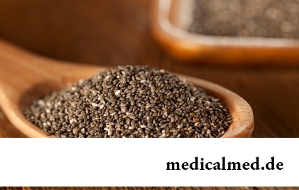
The chia plant, or the Spanish sage, is from South America. The indigenous people of the continent since ancient times used its seeds in food: small, but very nutritious kernels, in a form the reminding fasolina. Indians knew about useful properties of seeds of a chia, and applied...
Section: Articles about health
The next flu epidemic leads to the next panic, from year to year we give in on these manipulations: professionally alarming voice of the announcer in news, reports with calculation of the died patients, an interview with people in white dressing gowns and advertizing of anti-influenza means of different degree of inefficiency. All this reminds the Hollywood movies of epidemics threatening to destroy our planet. However, there is also one more similarity to cinema: everything comes to an end well. So, how to deal with the events, not in...
Section: Articles about health
Let's begin with the fact that a separate illness which is called "adjournment of salts", just does not exist. In practice this household name of plank beds...
Section: Articles about health
A lot of things depend on a condition of a backbone in a human body, a backbone - not only a support for a body, it also a receptacle for a spinal cord, that is why malfunctions with a backbone are so dangerous. To treat rachis diseases very difficult and long...
Section: Articles about health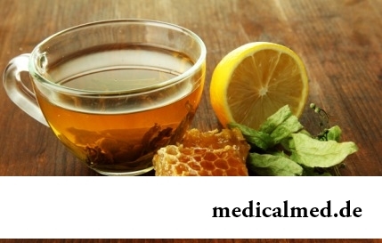
Cold – a state known to everyone which is followed by cold, cough, high temperature, a pharyngalgia. Often the first that we begin to do in hope again to become healthy – to accept medicines which are not always harmless whereas it is easy to facilitate displays of a disease by means of natural means. They not only softly eliminate disease symptoms, but also enrich the weakened organism with useful substances. We present you 8 drinks which are successfully used for...
Section: Articles about health
Eyes – one of the most vulnerable areas on a face therefore age changes concern them first of all. Whether it is possible to keep a pier...
Section: Articles about health
Nightmares belong to the most unpleasant frustration. Statistically, they happen at 4% of adults, and almost at 70% of children and teenagers. During a nightmare of people dreams himself in extremely difficult, life-threatening situation. It wakens suddenly, in...
Section: Articles about health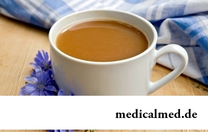
Coffee - the tonic loved by many for the invigorating aroma and deep taste. Having the stimulating effect, coffee increases working capacity, promotes concentration of attention, fights against drowsiness and improves mood. Statistically, about 30% of inhabitants of the planet regularly use coffee, from them more than 8% are "coffee-achievers" - the persons using more than 3 cups of drink a day....
Section: Articles about health
Aspirin (acetylsalicylic acid) – one of those drugs which are known literally to all. It is available in each home first-aid kit...
Section: Articles about health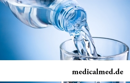
It would seem, about it there can be no disagreements: water is necessary for a human body for normal life activity, and about how and when it should be drunk, all know. It turned out that the situation is not absolutely so: for many years occur ве...
Section: Articles about health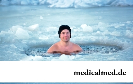
The winter swimming in open reservoirs called in our country by "winter swimming" – officially recognized sport and one of the most extreme ways of a hardening of an organism. This occupation has an old story and adherents in many countries. The international competitions in winter heats on open water, and every two years – the World Cup are annually held. Despite huge popularity and the proved advantage for health, winter swimming is still surrounded with hardy delusions. Ра...
Section: Articles about health