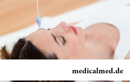
Endoscopy
 Endoscopy – diagnosis of internals by means of special devices – endoscopes.
Endoscopy method
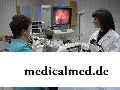
The technique of an endoscopic research is that through openings the soft tube on which end the lighting fixture and the microcamera are attached is entered into a body of the person. This tube is called the endoscope. Its diameter does not exceed 4 mm.
Different endoscopes are intended for the different fields of medicine. For endoscopy of a stomach, upper parts of a digestive tract, a 12-perstny gut, gastroduodenoskopa serve, for survey of a small bowel use enteroskopa, apply colonoscopes to endoscopy of intestines, use bronchoscopes to respiratory tracts.
At one manipulations the endoscope is entered through a mouth (endoscopy of a stomach), at others through a rectum (endoscopy of intestines), through a throat, an urethra and a nose (endoscopy of a nasopharynx). For carrying out, for example, a laparoscopy, in an abdominal cavity it is necessary to make special openings.
Types
There is a set of types of an endoscopic research. By means of this procedure it is possible to study a condition of such vitals as an abdominal cavity, a vagina, a small and 12-perstny bowel, ureters, bilious channels, a gullet, acoustic organs, bronchial tubes, a cavity of the uterus, and also to make endoscopy of a stomach, endoscopy of intestines, endoscopy of a nasopharynx.
The endoscope can be passed through vessels and to check their state, and also to see heart and heart cameras. In our century the endoscope can make the way even in a brain and give to the doctor the chance to see brain ventricles.
All types of endoscopic researches are directed to revealing the minimum changes of a mucous membrane which can lead further to oncology. Also the procedure allows to reveal oncology at an early stage and to remove a tumor that considerably increases chances of cancer patients of survival.
Cancer at early stages in general cannot be revealed by means of other research so there is no alternative of endoscopy today.
In addition to diagnosis, this procedure found broad application in surgery, urology, gynecology and other areas. With its help doctors stop bleedings, delete tumors at early stages. The procedure allows not only to carry out diagnosis of internals, but also to take a new growth fabric sample on the analysis.
Technique endoscopy of a forehead and eyebrows will also widely be applied in plastic surgery, for example. Endoscopy of a forehead allows to raise eyebrows, to remove or reduce quantity of mimic wrinkles on a forehead and between eyebrows. Endoscopy of a forehead enjoys wide popularity thanks to what practically does not leave hems.
As the procedure of endoscopy is carried out
At endoscopy of a stomach the device is entered through a mouth and on the monitor examine a mucous membrane. At the same time via the endoscope air supply is carried out – it is necessary for more detailed survey. The procedure lasts about 15-20 minutes.
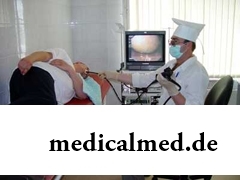 That the research was more exact, it is necessary to be prepared for it correctly. In 8-12 hours prior to the procedure it is desirable to eat nothing and not to drink.
Gastroscopy – the painful research causing an emetic reflex in the patient.
Transnasal endoscopy is transferred by patients much easier as the emetic reflex at it is absent.
Endoscopy of a stomach is done for specification of the diagnosis and identification of changes.
Performing endoscopy of intestines – more painful and long occupation. Pains can be caused by features of a gut, commissures. The procedure takes 30 minutes till 1 o'clock. Often when carrying out a kolonoskopiya use an anesthesia.
When carrying out a kolonoskopiya preparation is also important. Here it is recommended three days prior to the procedure to pass to a bezshlakovy diet.
Indications to a kolonoskopiya are disturbances of a chair, mucifying and blood, painful feelings, bleeding from a large intestine.
Bronkhoskopiya is led by introduction of the thin endoscope through a nose, a throat and phonatory bands directly to a trachea. It allows to examine a bronchial tree from within. The research is shown at pneumonia, bronchitis, suspicions of a tumor.
At endoscopy of a nasopharynx the endoscope which allows to see a picture in a nose and possible polyps is entered into a nose. Endoscopy of a nasopharynx is shown at the complicated breath, nasal bleedings, disturbance of sense of smell, at polyps and not clear headaches.
Endoscopy of a nasopharynx allows to reveal pathological changes in mucous a nose without intervention of surgical methods.
Video capsular endoscopy
This look is the new direction in medicine. The method is that the patient swallows of the plastic capsule which in size is no more than the ordinary capsule with medicine. The capsule passes on all digestive organs, at the same time all image is fixed on the special device, and it, in turn, transfers all data to the screen.
Video capsular endoscopy was patented in America at the beginning of this century and in high gear gains steam. The capsule weighs 4 гр., and length makes it 2,5 cm. One end at the capsule transparent, behind it the lens, the microcamera and LEDs is hidden. In other part of the capsule there are a transmitter, the battery and the antenna.
Video capsular endoscopy is very convenient as it allows without separation from major activities of the patient to carry out full endoscopy of a stomach, endoscopy of intestines and digestive tract. Besides, the similar research allows to see even those sections of intestines which are not available when carrying out an ordinary endoscopic research.
However video capsular endoscopy has one essential minus. Unfortunately, by means of this technique it is possible to conduct only a research of digestive organs.
The liver is the heaviest body in our body. Its average weight makes 1,5 kg.

Statistically, pathologies of a thyroid gland in the world more than 500 million people have. Failures in work of this body conduct to is heavy...
Section: Articles about health
Ability of an organism to resist to adverse environmental factors (to impact of temperature drops, humidity and pressure, to the attacks of causative organisms, etc.) directly depends on what the person eats. Business here not only in, that C...
Section: Articles about health
Cold – a state known to everyone which is followed by cold, cough, high temperature, a pharyngalgia. Often the first that we begin to do in hope again to become healthy – to accept medicines which are not always harmless whereas it is easy to facilitate displays of a disease by means of natural means. They not only softly eliminate disease symptoms, but also enrich the weakened organism with useful substances. We present you 8 drinks which are successfully used for...
Section: Articles about health
Diapers for adults – individual one-time means of hygiene which in some situations is irreplaceable, and from such situats...
Section: Articles about health
About influence of fasting days on an organism it is told much – both about advantages, and about shortcomings. It is considered that fasting day in the form of a short-term monodiet is useful, promoting effective removal of slags from an organism whereas irregular, it is excessive п...
Section: Articles about health
Producers of milk mixes for children assure: mixes are ideally balanced and adapted for needs of babies. If mother should raise artificially the kid owing to serious problems with health, to do nothing – it is necessary to feed with substitutes of milk. However pediatricians note that not seldom women without good reasons refuse feeding of the child a breast and pass to milk mixes. Common causes of such decision – the aspiration to leave quicker...
Section: Articles about health
Wood louse – the ordinary-looking unpretentious plant extended in all territory of our country. It quickly expands, and sometimes for...
Section: Articles about health
New year, wedding, birthday, office party – an occasion to drink at the Russian person will always be. How to reduce a negative impact of alcohol by an organism and to avoid a condition of strong intoxication? The most correct council – to refuse the use spirits напитк...
Section: Articles about health
The summer of this year in Russia was very ambiguous. Regions suffered from a merciless heat, from pouring rains, the hail from time to time dropped out, then there was again a heat which alternated with rainfall again. Many people suffer from such sharp changes of weather. Even flu epidemics and a SARS were recorded....
Section: Articles about health
Helminthosis is one of the most widespread diseases. Statistically, with any species of helminths it is infected porridges...
Section: Articles about health
The number of long-livers is very small. One person from 5 thousand lives up to age of 90 years, and the centenary boundary steps over only one of 20 thousand. However, doctors claim that each of us is quite able to affect own destiny. At the same time speech to Ida...
Section: Articles about health
Stroke (acute disorder of cerebral circulation) – one of the most widespread neurologic diseases. Annually in the world more than 6 million people die of this illness. From the survived patients about 80% become disabled people, and nearly a third from them needs afterwards permanent care. In fact, the stroke creates a situation at which a part of cells of a brain loses blood access, loses an opportunity to receive oxygen and nutrients, and perishes. As a result of a razviv...
Section: Articles about health
Among a set of the perfumery and cosmetic goods which are released today the special group is made by the means containing anti-bacterial...
Section: Articles about health
What they, women? Beautiful, gentle, passionate and at the same time windy, gusty, and nervous. And what is stranger: have all these qualities of the woman at the same time. But here only the mood their time sharply changes on completely opposite: in the morning...
Section: Articles about health
Epilepsy is one of widespread neurologic diseases. Parents, whose children suffer from this illness, should face rumors and delusions, many of which remained since the Middle Ages....
Section: Articles about health
Many parents of children at the age of 2-4 years face excessively whimsical behavior of the child. The kid exhausts constant crying...
Section: Slideshow
All got used long ago that, having addressed the plastic surgeon, it is possible to modify natural parameters of a figure or to minimize the damages put to appearance with ruthless time. Many people (preferential women) worldwide е...
Section: Articles about health
Climax - process of fading of reproductive function of an organism in process of its aging. At women the main sign of its approach is the termination of a menstrual cycle. Officially the menopause is diagnosed when periods are not observed within 12 months. Age changes quite often are followed by emotional failures, disturbance of thermal control and sweating, dizzinesses and headaches, tachycardia and other unpleasant phenomena. This complex of symptoms...
Section: Articles about health
Such trouble as the milkwoman's attack, at least once in life happened almost to each woman. Prevalence забол...
Section: Articles about health
Cold, puffiness of a nose, itch, the watering eyes - characteristic symptoms of the allergic rhinitis resulting from hit of allergens (pollen, house dust, hair of animals, etc.) on a mucous membrane of a nose. Unpleasant feelings often deliver беспоко...
Section: Articles about health
So, you resolved to lose weight. And now you try to understand what to begin with: from exercise stresses or a diet? And how to make that process of weight loss did not give you an inconvenience, and, on the contrary, brought joy?...
Section: Slideshow
The sudden heat on all body which is followed by perspiration and a cardiopalmus – the phenomenon familiar to many people. Most often t...
Section: Articles about health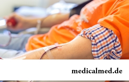
Transfusion of donor blood has almost century history. In spite of the fact that this procedure is quite usual for many people, process of blood donation is still surrounded with numerous myths. Today we aimed to discredit the most widespread of them....
Section: Articles about health
"Epilepsy" doctors made the diagnosis in antique times. Displays of an illness and pattern of its development are very well studied. However for nonspecialists this disease remains to not less mysterious, than in the ancient time. Many delusions are connected with epilepsy, and it sometimes very unpleasantly affects quality of life of patients and their relatives. In this article we will try to dispel the most known of similar myths....
Section: Articles about health
Women quite often suffer from complexes concerning the sizes of the bust. Strangely enough, reason душевног...
Section: Articles about health
Proofs of efficiency of Mildronate at treatment of coronary heart disease with stenocardia can be found in many publications of the end of the twentieth century. Researches were conducted since 1984, including placebo - controlled effects. In total клиничес...
Section: Articles about health
Feeding by a breast - the integral part of ideal motherhood allowing to come into contact with the kid and to create to it healthy immunity since early years. Nevertheless, this important process in life of mother and child can be saddened laktostazy − by a milk delay in a mammary gland. What main reasons for a laktostaz? How not to allow problems with breastfeeding? Let's consider 10 premises resulting in stagnation of milk at the nursing mother....
Section: Articles about health