Diplopia
General characteristic of a disease
The diplopia is the ophthalmologic pathology connected with sight doubling. The objects which are coming into the view of the person look double as a result of a deviation of an axis of one of eyes. A number of the reasons of ophthalmologic, neurologic or infectious character can cause similar disturbances.
Diplopia reasons
Development of a diplopia can provoke shift in an eyeglobe eye-socket. Quite often bring eye injuries to it, for example, the infringement of eye muscles caused by an eye-socket wall change. The abnormal provision of an eyeglobe is caused also by hematomas of eye fabrics.
Other possible reason of a diplopia - damage of a third cranial nerve. Aneurism of a carotid artery, intracranial tumors or meningitis of a tubercular etiology can provoke it.
Also infectious processes which affected a brain trunk at a rubella, parotitis, diphtheria or tetanus are the reasons of a diplopia. Also severe alcoholic or medicinal intoxication can cause a diplopia.
Doubling of the image or diplopia is quite often observed against the background of botulism, a thyrotoxicosis, multiple sclerosis or hysterical attacks. The diplopia reason as one of types of postoperative complications, manipulations can serve in the eyes at surgical treatment of amotio of a retina, squint or cataract.
Diplopia symptoms
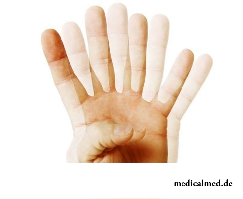
The main complaint of patients with a diplopia – doubling of the image. In most cases doubling of objects of surrounding reality happens at sight two eyes. The diplopia binocular is so demonstrated. Doubling of sight can be partial and be displayed only in a certain zone of a field of vision or full. Manifestation of a diplopia also individually depending on remoteness of the considered objects. In certain cases doubling happens only at a look on close located or, on the contrary, is exclusive on remote objects.
Two images of one subject arising at a diplopia have the different brightness and contrast. One of them is usually a little displaced down, and also across and located under a certain corner to the second image.
Because of the progressing diplopia the patient loses labor skills. To it can be heavy to perform homework, to manage transport, and sometimes and just to move. To return image sharpness to the person with a binocular diplopia it is necessary to close one of eyes. The patient with other type of a disease, a diplopia monocular, such measure does not help.
Types of a diplopia
Diplopia binocular - the most common form of the doubled sight. At a binocular diplopia the visual picture of both eyes is not projected in the corresponding retina points. The visual axis moves, and the patient with a binocular diplopia sees the doubled image of objects. The binocular diplopia can be the motor, touch or mixed, constant or temporary, neuroparalytic, orbital, caused injuries, strabogenny at squint, etc.
Monocular diplopia – more rare pathology of the doubled sight. Disturbances of the image in this case arise even at sight one eye. The image projection at the same time to two different points of a retina of one eye leads to a monocular diplopia. The monocular diplopia is caused most often by an incomplete dislocation or a partial phacoscotasmus. The reason of a monocular diplopia can appear also an iridodialysis (a separation of an iris of the eye from a ciliate body as a result of an eye injury) or a polycoria (inborn pathology of a structure of an iris with several openings).
Diagnosis of a diplopia
Primary diagnosis of a diplopia is established on the basis of complaints of the patient concerning doubling of images. Further diagnosis of a disease continues by means of test control over sight of the person whose look is directed to the moving light source.
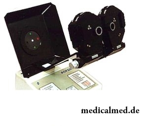
By means of mapping of coordinates of the received images the doctor manages to diagnose what of eye muscles is struck. More modern method of definition of the injured extraocular muscle of an eye – a koordimetriya by means of an oftalmokoordimetr OK.
Diagnosis of a diplopia assumes also obligatory assessment of a condition of situation and mobility a century with the help the Cover test. Inspection of a conjunctiva of eyeglobes is in addition performed, visual acuity, a refraction and color sensation is checked.
Treatment of a diplopia
Treatment of a diplopia of secondary type of a neurologic, infectious or ophthalmologic etiology assumes therapy of the main disease.
Treatment of a diplopia as a basic disease is under authority of the neuropathologist or neurosurgeon. The surgeon-ophthalmologist who is carrying out a resection or plastics of eye muscles takes part in treatment of a diplopia of the traumatic nature. At the same time to perform eye muscle operation it is allowed, as a rule, only 6 months later after an injury.
Optical correction of a diplopia is carried out by means of a prismatic spectacles. They are capable to improve the clearness of sight of the patient significantly. Optimum correction in treatment of a diplopia makes 6 prismatic dioptries on each eye.
Glasses wearing with bigger prismatic compensation is in rare instances allowed. Frenelevy prisms can be valid to 20 ave. of dioptries, however already at compensation 15 ave. of dioptries they influence visual acuity and create effect of an iris of the eye around visible objects.
Functional treatment of a diplopia consists in performance of special exercises across Kashchenko for recovery of abilities of solid vision and expansion of a field of vision, and also exercise on merge of objects by means of red glass, etc.
In Great Britain there is a law according to which the surgeon can refuse to do to the patient operation if he smokes or has excess weight. The person has to refuse addictions, and then, perhaps, he will not need an operative measure.

The body of the person almost for 60% consists of water. It is so important for normal functioning of an organism that loss of all is ponut...
Section: Articles about health
Each person knows that fervescence is an illness sign. However too low temperature (hypothermia), especially also can demonstrate existence of diseases when it is observed long enough. Such state is dangerous those...
Section: Articles about health
It is known that the person for 80% consists of water which participates in all processes of an organism. The person loses liquid daily – as a result of sweating, breath, an urination, and its insufficient completion due to various reasons can lead to dehydration of varying severity. Dehydration (dehydration) occurs already in case of loss of liquid in number of 1% of body weight and can result both in easy thirst, and by the death. In time to notice signs обезвож...
Section: Articles about health
The number of long-livers is very small. One person from 5 thousand lives up to age of 90 years, and the centenary boundary steps only about...
Section: Articles about health
Radiological methods of a research are applied in medicine more than hundred years, and thanks to them millions of lives were saved. In many cases without X-ray it is impossible to make exact idea of a condition of bodies and fabrics, it is correct to make the diagnosis. Those...
Section: Articles about health
EKO, or extracorporal fertilization - a method of treatment of infertility which became the reason of a set of broken-down copies in due time accused the people working on its creation neither more nor less of rivalry good luck. Already very few people deny the right of a method for existence, and to surprise nobody with "children from a test tube". And nevertheless, a certain magic in the procedure of artificial fertilization is, process of origin of new life is always a secret, and even it р now...
Section: Articles about health
All know that self-treatment is dangerous. However absolutely it is almost impossible to do without it. Rate of modern life not on...
Section: Articles about health
Visit of doctors – business not the most pleasant, and many people do not hurry to undergo necessary planned inspections. Such behavior is extremely thoughtless and improvident. Our health is necessary not only to us: wellbeing of darlings, children, grandsons and престар...
Section: Articles about health
It would seem, to buy drugs in Moscow does not make a problem – a drugstore, and not one, is available for each resident of the capital within walking distance. And, nevertheless, Internet drugstores become more popular – what it is possible to explain such phenomenon with? Actually there is a lot of reasons and if to formulate them it is short, then the most suitable word will be - "conveniently". We suggest to get acquainted in more detail with pluses and minuses of online drugstores that buying drugs, not to make the wrong choice....
Section: Articles about health
On health of the person physicians know about salutary action of animals long ago. About 7 thousand years ago great Hippocrates рекоменд...
Section: Articles about health
Many parents of children at the age of 2-4 years face excessively whimsical behavior of the child. The kid exhausts constant crying and whims not only the parents, but also himself. In what the reasons of children's whims. And how to fight with them?...
Section: Slideshow
Summer in the heat. Many are going to spend vacation abroad. Travelers the tender seas, rest on beaches wait, for sightseeing, campaigns on natural and cultural reserves. But, unfortunately, on vacation also problems with health can wait for us. On a foreign trip it is possible to face also diseases which not only will spoil long-awaited issue, but also will force to be treated within long months after its termination. To be insured completely from troubles of it a sort...
Section: Articles about health
Dogrose – one of the most widespread adornment and medicinal plants growing practically in all territory ours...
Section: Articles about health
Very often as a source of the infection which caused a disease serves our house - the place which a priori has to be safe. However disease-producing bacteria can perfectly feel not only in insanitary conditions, but also in our apartment if not осущ...
Section: Articles about health
The pancreas performs two functions in a human body: release of enzymes without which digestion of carbohydrates, fats and proteins, and a producing hormones is impossible. The most important of them - insulin, is the main participant of carbohydrate metabolism normalizing processes of education and utilization of glucose, the main energy source for an organism....
Section: Articles about health
The phenomenon of improvement of a condition of the patients at administration of drugs who are not containing active agents, so-called effect of placebo is known...
Section: Articles about health
Life expectancy in various regions of Earth is not identical. Social stability, economic wellbeing, availability and level of medical care, household comfort, literacy of the population in the field of observance sanitary гигиен exert impact on it...
Section: Articles about health
Epilepsy is one of widespread neurologic diseases. Parents, whose children suffer from this illness, should face rumors and delusions, many of which remained since the Middle Ages....
Section: Articles about health
Tick-borne encephalitis – one of the most dangerous viral diseases which causative agents transfer and is given to people by ixodic mites. Эт...
Section: Articles about health
The pine is one of the most widespread plants of our woods. Its needles and pitch not without reason called by "gallipot" were since ancient times used for strengthening of protective forces of an organism, treatment of avitaminosis, anemia and many other diseases. In recent years wide п...
Section: Articles about health
The state of health of the person depends on many factors. One of the most important is the constant, but not exhausting a physical activity. In the presence of various illnesses specialists often advise patients to do swimming which by right takes the leading place by efficiency of improvement, having at the same time a few contraindications. Today we will talk about the main directions of therapeutic impact of swimming on a human body....
Section: Articles about health
Sooner or later hair turn gray at all. Many people try to hide these changes, returning natural color of the hair with the help about...
Section: Articles about health
About 10-15 years ago existence of the computer in the apartment of the Russian was considered as a rarity and office rooms were only at the first stage of equipment by these useful devices. Today practically in each house there is a computer (and often not one), and to constants...
Section: Articles about health
The hysteromyoma is diagnosed more than at a third of women 35 years are more senior. This high-quality new growth which at early stages successfully resolves by means of medicines. It is necessary to resort to an operative measure only when patients too late address specialists, or therapeutic methods do not give the expected effect because of specific features of an organism. Besides, there is a large number of quite effective national...
Section: Articles about health
For the city dweller the fitness is the most convenient sport. It is enough to acquire the subscription to the gym to receive to a toast...
Section: Articles about health
Statistically, pathologies of a thyroid gland in the world more than 500 million people have. Failures in work of this body lead to heavy disbolism, development of heart diseases, vessels, a reproductive and nervous system. In hard cases excessive...
Section: Articles about health
Statistically, in Russia about 34% of citizens smoke. Most of consumers of tobacco has problems with health sooner or later. Not only smokers, but also their relatives suffer. Besides, cigarettes are expensive, and need of their acquisition goes a heavy burden on the budget of thousands of Russian families. Many people dream to refuse harmful tendency, but everyone manages to make it not: nicotine addiction is affectionate and to get rid of it not easy....
Section: Articles about health