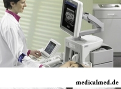


Ultrasonography of mammary glands
 Ultrasonography of mammary glands – a painless, safe and informative way of diagnosis of a breast with drug ultrasonography use.
Ultrasonography of a breast becomes for the purpose of diagnosis of various volume educations found at a palpation. It is considered that ultrasonography successfully supplements mammography. By means of an aim puncture biopsy under ultrasonic control it is possible to achieve more exact assessment of pathology.
When it is necessary to make ultrasonography of mammary glands
- At pregnancy and in the period of a lactation;
- At complaints to diseases of a mammary gland;
- For the purpose of routine inspection of mammary glands, an onkoosmotr (for women 30 years – once a year, for women after 30 years – 2 times a year are younger);
- At inflammatory process in dynamics;
- For specification of the diagnosis at identification of new growths of mammary glands at a palpation or at x-ray mammography;
- At a nodal mastopathy with atypical manifestations;
- At axillary lymph nodes, for the purpose of clarification of the nature of their increase;
- For diagnosis of cysts of various size;
- For assessment of a condition of silicone prostheses of mammary glands;
- At a simultaneous sklerozirovaniye of several cysts.
Advantages and shortcomings
- To make ultrasonography of mammary glands, no injections and needles therefore the procedure absolutely painless will be required;
- Drugs ultrasonography are readily available, is practically in each hospital, and the research does not take a lot of time cheaper than other ways of diagnosis;
- Interpretation of ultrasonography of mammary glands can be used for demonstration in real time that is convenient when holding additional procedures, in particular, to a biopsy;
- Ultrasonography is used for detection of pathologies which are difficult for receiving when carrying out only one mammography;
- It is possible to define by ultrasonography not only sick zones, but also healthy.
When performing ultrasonography of mammary glands interpretation of result sometimes is a problem. Interpretation of ultrasonography of mammary glands can demand carrying out a number of additional researches in the form of a biopsy or ultrasound. The majority of suspicious zones as a result of additional researches are healthy;
Preparation for ultrasonography of mammary glands
To carry out ultrasonography of mammary glands, preparation is not necessary. Specialists recommend to conduct a research from the 5th to the 14th day of a menstrual cycle.
Technology of carrying out research
The patient ask to undress above a belt. Most often the research is conducted in a dorsal decubitus. The doctor will apply on the woman's breast special gel on a water basis then will put the transmitter closely to a body, smoothly moving it on a target zone before obtaining necessary results. In most cases pressure of the transmitter does not cause any discomfort.
 First of all the healthy breast looks round, and then conduct gland researches with probable pathology. The research includes obligatory scanning of four zones of lymph nodes: supraclavicular, subclavial, axillary and rebreast zones. All procedure takes no more than 10-15 minutes.
Contraindications to a research
Ultrasonography – the harmless noninvasive procedure therefore there are no contraindications to its carrying out.
Special instructions
- Ultrasonography can be used for diagnosis of mammary glands, but it is not the basis for refusal of visit of the mammologist;
- The set of cancer tumors cannot be seen, having made ultrasonography of mammary glands. For this purpose the biopsy needs carrying out a number of additional researches, in particular;
- After carrying out a biopsy the majority of focal zones are healthy;
- By preparation for ultrasonography of mammary glands it is important to choose hospital which specialization is ultrasonography of a breast as there more narrow-purpose specialists work;
- Interpretation of ultrasonography of mammary glands depends on whether the doctor who is carrying out the procedure, pathology during primary check will notice. And experience and the high-quality equipment is for this purpose important.
Having fallen from a donkey, you more likely will kill yourself, than having fallen from a horse. Only do not try to disprove this statement.

Life activity of one-celled fungi of the sort Candida is a proximate cause of development of candidiasis (milkwoman), it is related...
Section: Articles about health
For residents of the countries of Southeast Asia various algas are an obligatory component of a daily diet. Their popularity is connected not only with high tastes, but also with numerous curative properties. Russians are a little familiar with...
Section: Articles about health
Smack in a mouth can arise in the natural way – as a result of lack of morning hygiene or reception of the corresponding food. However in certain cases its existence is a sign of certain pathologies, and allows to reveal an illness at an early stage. Depending on character of aftertaste – acid, salty, bitter, sweet – distinguish also diseases which accompany it....
Section: Articles about health
The brain of the person is studied not one hundred years, but the quantity of the riddles connected with this body increases rather, than reducing...
Section: Articles about health
For the help to doctors in the choice of optimal solutions for treatment of various diseases the Cochrane scientific organization (Cochrane) conducts joint researches with representatives of scientific community around the world. The analysis of a series became carried out by one of the last methanolyses...
Section: Articles about health
Very often as a source of the infection which caused a disease serves our house - the place which a priori has to be safe. However disease-producing bacteria can perfectly feel not only in insanitary conditions, but also in our apartment if not to carry out due care of favourite places of their dwelling. What they − sources of their reproduction? Let's consider 10 most widespread places in our house, the most dangerous from the point of view of infection with microorganisms....
Section: Articles about health
The unpleasant feelings connected with spring breakdown are familiar almost to each of us. Often happens that in March-April on the person...
Section: Articles about health
It would seem, to buy drugs in Moscow does not make a problem – a drugstore, and not one, is available for each resident of the capital within walking distance. And, nevertheless, Internet drugstores become more popular – what it is possible to explain such phenomenon with? Actually m reasons...
Section: Articles about health
Dark circles (bruises) under eyes – a shortcoming with most of which often fight against the help of cosmetics (proofreaders, saloon procedures and so forth), eliminating only its visibility. However, according to doctors, skin around eyes – the indicator of many disturbances in an organism. To reveal them at early stages, without having disguised bruise, and having addressed its reasons – a task of each person who is regularly finding under with own eyes dark stains. Early detection and elimination of the disease lying in wasps...
Section: Articles about health
Partial and the more so full loss of hearing significantly reduces quality of life. Difficulties with communication lead to loneliness and замкн...
Section: Articles about health
Diapers for adults – individual one-time means of hygiene which in some situations is irreplaceable and from such situations any person is not insured. Though nobody perceives need of their use with enthusiasm, however without it to a sra...
Section: Articles about health
With age in a human body harmful substances collect. We receive them with food and water, at inhalation of the contaminated air, reception of medicines, use of household chemicals and cosmetics. A considerable part of toxins accumulates in a liver which main function is continuous purification of blood. This body begins to knock as any got littered filter, and efficiency of its work decreases....
Section: Articles about health
Is told about advantage of domestic animals for development of the child much. But many parents nevertheless do not hurry to bring pets as about...
Section: Articles about health
Kidneys perform the most important function of clarification of blood from those products of metabolic processes which cannot be used by an organism for obtaining energy and construction of new cells. With the urine produced by kidneys from a body of the person bulk is removed...
Section: Articles about health
Smoking not only exerts a negative impact on the state of health of the consumer of tobacco products, but is an air polluter the substances potentially dangerous to people around. In recent years significantly the number of the people aiming to get rid of an addiction increased. Business this difficult: having left off smoking, the person immediately begins to suffer from abstinence. Besides, many yesterday's smokers feel at first great disappointment as улучш...
Section: Articles about health
The fatigue, sleep debt, disturbances of food, bad mood, vagaries of the weather – all these circumstances badly are reflected in our vn...
Section: Articles about health
High temperature - a frequent symptom of such widespread diseases as a SARS, quinsy, pneumonia, etc. To reduce heat, having facilitated a condition of the patient, doctors recommend to accept antipyretics, however their use is not always possible. Too h...
Section: Articles about health
Practically each person is familiar with the annoying, pulling, unscrewing pains caused by overcooling of muscles of a back. In certain cases inflammatory process is not limited to discomfort, being followed by emergence of hypostasis, consolidations, temperature increase. At the wrong treatment the acute miositis can lead to a chronic disease or aggravation of other pathologies of a back (vertebral hernia, osteochondrosis) therefore it is important to pay attention to symptoms of an illness in time and to start to...
Section: Articles about health
The phenomenon of improvement of a condition of the patients at administration of drugs who are not containing active agents, so-called effect of placebo is known...
Section: Articles about health
The varicosity has familiarly many, statistically, this disease more than a half of all adult population. As a rule, the varicosis affects preferential superficial vessels, and is shown by characteristic cosmetic defects. Guo...
Section: Articles about health
Proofs of efficiency of Mildronate at treatment of coronary heart disease with stenocardia can be found in many publications of the end of the twentieth century. Researches were conducted since 1984, including placebo - controlled effects. In total clinical tests of Mildronate were carried out for more than thirty years....
Section: Articles about health
About 10-15 years ago existence of the computer in the apartment of the Russian was considered as a rarity and office rooms were only on перв...
Section: Articles about health
Bees – really unique beings. Practically all products of their life activity are used by the person. Since the most ancient times medicinal properties of honey and other substances received in the course of beekeeping are known. The fact that all these пр is especially significant...
Section: Articles about health
An eye of the person daily experiences considerable strain. The problem of preservation of sight is for many years directly connected with a question of supply of tissues of eye enough oxygen and nutrients. This task is carried out by small vessels – capillaries. For normal functioning of the visual device extremely important that they kept the integrity, but it works well not always. Microtraumas of eye vessels during which there are small hemorrhages it is extraordinary расп...
Section: Articles about health
At this plant there are a lot of names: tuberiferous sunflower, Jerusalem artichoke, solar root, earth pear. Contrary to spread...
Section: Articles about health
All like to sing. Small children with pleasure are engaged in a vocal, not especially thinking of hit in a melody. Adults most often hesitate, being afraid to show lack of talents in this area, and it is vain: singing is very useful for health....
Section: Articles about health
Tick-borne encephalitis – one of the most dangerous viral diseases which causative agents transfer and is given to people by ixodic mites. These are the small blood-sicking insects living in the considerable territory of our country. The person bitten by a tick can catch also erlikhiozy, bartonnelezy, babeziozy, mycoplasmosis and Lyme's disease. As well as encephalitis, these illnesses affect the central nervous system, and as specific antiviral therapy does not exist, the forecast very to a neuta...
Section: Articles about health
