Tibia
Tibia – a large and long bone of a shin. The bone consists of a body and two epiphysis – lower distal and upper proximal.
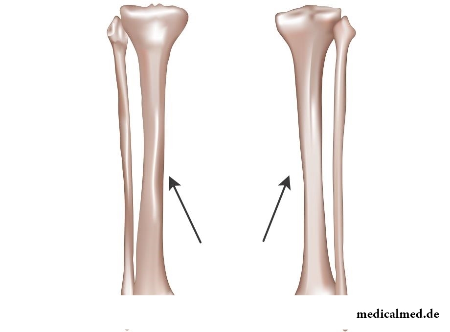
Structure of tibia
The body of a bone has the trihedral form with three edges – front, medial and interosseous, and three surfaces – medial, back and lateral.
The first line of a bone has the pointed form and reminds by the form a crest. In an upper part it turns into tuberosity. The interosseous edge has the pointed form and an appearance of a comb. This comb is directed towards a fibular bone. The medial surface of a bone slightly convex is also well probed through skin together with a first line of a shaft of the tibia.
Lateral (anteroexternal) surface of a bone slightly concave. And the back surface has the flat form. On a back surface there is a line of a soleus muscle which extends from an ectocondyle medially and down. A little from below the nutritious opening which lasts in distally the directed nutrient canal is located.
The proximal epiphysis of tibia is a little expanded. Its side parts are lateral and medial condyles. Outside of an ectocondyle the flat fibular joint surface is located. At the top of a proximal epiphysis on average department there is an intercondyloid eminence in which it is possible to distinguish two hillocks:
- internal medial intercondyloid behind which it is possible to distinguish the back intercondyloid field;
- outside lateral intercondyloid in front of which the front intercondyloid field is located.
Two fields are the place of fastening of crucial knee ligaments. On each side an intercondyloid eminence on an upper joint surface the joint surfaces having the concave form – medial and lateral reach for each condyle. Concave joint surfaces are limited on the periphery to edge of a tibial bone.
The distal epiphysis of a bone has a squared shape. On its lateral surface there is a fibular cutting adjacent to a distal epiphysis of a fibular bone. On a back surface of a distal epiphysis there passes the lodyzhkovy furrow. In front from a furrow the medial edge of a distal epiphysis of a tibial bone passes into a medial anklebone – the shoot directed down which is well palpated. On the lateral surface of an anklebone the joint surface of an anklebone is located. It passes into the lower surface of a bone and lasts in the lower concave joint surface of a tibial bone.
Fracture of tibia
All fractures of tibia share on:
- slanting;
- cross;
- intra joint;
- fragmentary;
- splintered.
Fractures of a medial anklebone and condyles of a tibial bone belong to intra joint changes. The medial anklebone serves as the internal bone stabilizer of an ankle joint. As a rule, its change results from twisting of a shin with the fixed foot. Also often the fracture of an internal anklebone results from not physiological sharp turn of foot.
Main symptoms of fractures of tibial bone:
- Tibia at the movement and a palpation hurts;
- Because of the shift of bone fragments the shin is deformed (the extremity axis changes);
- There is hypostasis;
- It is impossible to carry out axial load of a leg.
Treatment of changes is preferential performed by means of an operative measure. As a rule, the patient can carry out load of a sore leg the next days after operation.
Cyst of tibia
Quite often, when tibia hurts, it can demonstrate existence of a cyst.
Bone cyst – a disease during which the thickening in a cavity of a bone tissue is formed.
Still the origin of cysts of bones is definitely not found out. It is established that cysts of tibia develop as a result of frustration of a hemodynamics on the limited site of a bone. In fact, formation of a cyst is dystrophic process. Disturbance of intra bone blood circulation and activation of the lysosomic enzymes leading to destruction of collagen, glucosaminoglycans and other proteins is the cornerstone of formation of cysts. On the international classification of a cyst carry to opukholepodobny diseases.
The bone cyst can be solitary and anevrizmalny. The solitary cyst develops throughout a long span, meets at youthful age at males more often. The Anevrizmalny cyst arises suddenly and develops quickly. Most often, the anevrizmalny cyst results from a direct injury of a bone.
Despite the general nature of these diseases, they can be distinguished accurately as they have different symptoms and X-ray patterns.
Each person has not only unique fingerprints, but also language.

Deciding to get rid of an addiction, not all imagine what effects it is necessary to face. Process of refusal from ку...
Section: Articles about health
Bathing in broths of medical flowers and plants (phytobathtub) was eurysynusic since Cleopatra who is a good judge in all that concerns beauty and health. And today phytobathtubs is the simple and available means allowing not only to remove nervous N...
Section: Articles about health
For the last decades the diabetes mellitus of the second type became really world problem. The number of cases annually increases, and average age of patients for whom the illness is diagnosed, steadily decreases. Specialists consider that one of the main reasons for this trouble is disturbance of a diet. In other words, the huge number of people regularly overeats or excessively is fond of the products causing glucose exchange process failures....
Section: Articles about health
Transfusion of donor blood has almost century history. In spite of the fact that this procedure is quite usual for many people, itself п...
Section: Articles about health
Osteoporosis this general disease which main sign is decrease in density of a bone tissue. On distribution width it takes the fourth place among noninfectious diseases. The illness develops at mature age more often: in our country to them harvest seasons...
Section: Articles about health
Contrary to popular belief, the multiple sclerosis (MS) is not connected neither with sclerous changes of walls of vessels, nor with age forgetfulness and problems with concentration of attention. This disease has the autoimmune nature. Pathological process is expressed in degradation of nervous tissue and destruction of its enveloping layer - a myelin. Multiple damages of the central nervous system which are shown by decrease in sight, bystry fatigue, on become result of development of an illness...
Section: Articles about health
The name of this disease precisely reflects the problem reason: it consists in the bra fastener pressure upon a certain zone...
Section: Articles about health
Dietary supplements (dietary supplements) for the last decades were so thoroughly included into our life that, apparently, it is already impossible to find the person who at least once did not try them. At the same time, most of our compatriots have a vague idea about...
Section: Articles about health
Cystitis, or inflammation of a mucous membrane of a bladder, this very widespread disease which, owing to some features of a structure of bodies of urinogenital system, women have approximately four times more often than men. Women aged from 20 up to 45 years enter into the main risk group. Cystitis is an illness of a bacterial origin. It can have an acute or chronic current. The second option is dangerous not only a frequent recurrence, серьезн...
Section: Articles about health
Iodine - one of thirty most important microelements in our organism. The main role of iodine consists in synthesis thyroid гормо...
Section: Articles about health
Not everyone can brag of the shining Hollywood smile. Even the person who is regularly visiting the stomatologist and watching of oral cavities over health periodically has problems: enamel of teeth darkens under the influence of some products, on it I accumulate...
Section: Articles about health
Today about 30 diseases, sexually transmitted are known. Wide circulation of these illnesses is extremely promoted by the dual attitude towards them: on the one hand, most of people know about "shameful" diseases very little and do not aim at receiving detailed and reliable information, considering that such problems personally will never concern them. With another – there are delusions about STD which instill unreasonable confidence that troubles such...
Section: Articles about health
When overcomes feeling of hunger, and an opportunity to have dinner fully is absent, having a snack − small on volume comes to the rescue...
Section: Articles about health
What they, women? Beautiful, gentle, passionate and at the same time windy, gusty, and nervous. And what is stranger: have all these qualities of the woman at the same time. But here only the mood their time sharply changes on completely opposite: in the morning...
Section: Articles about health
Mushrooms - the surprising inhabitants of our planet having a set of wonderful qualities. Thanks to one of them, a mold mushroom of Penicillium notatum, the first natural antibiotic - penicillin was received nearly 80 years ago. The mankind is obliged to this opening by millions of saved lives....
Section: Articles about health
For the time being the perspective of heart diseases seems to most of people remote and foggy. But sooner or later практичес...
Section: Articles about health
EKO, or extracorporal fertilization - a method of treatment of infertility which became the reason of a set of broken-down copies in due time accused the people working on its creation neither more nor less of rivalry good luck. Already very few people deny the rights...
Section: Articles about health
The kid who was recently born is surrounded with love of adult family members and their cares without which the baby cannot exist. Some parents consider that gentle attachment and caress are quite enough that the child correctly developed and was happy, but it not so. It is important to know as much as possible about specifics of care of the baby, the reasons of his behavior and possible problems. Only the "able to see" love will provide to the little man that it is necessary for him....
Section: Articles about health
It is possible to find the extensive range of fruit and vegetables in modern shops. Russians already got used that on counters in any...
Section: Articles about health
The modern person not always manages to find housing in the environmentally friendly region and such work which would not do harm to health. With food stuffs at first sight the situation is much better: shops are overflowed with goods which are positioned пр...
Section: Articles about health
The winter swimming in open reservoirs called in our country by "winter swimming" – officially recognized sport and one of the most extreme ways of a hardening of an organism. This occupation has an old story and adherents in many countries. The international competitions in winter heats on open water, and every two years – the World Cup are annually held. Despite huge popularity and the proved advantage for health, winter swimming is still surrounded with hardy delusions. Ра...
Section: Articles about health
Food with the increased content of sugar is attractive to most of people - it is scientifically confirmed fact. Business here not in a nevozder...
Section: Articles about health
Household skills which to us so diligently imparted in the childhood it appears, not always bring only benefit. According to results of the last researches, some habits which for a long time were considered useful and even necessary can become...
Section: Articles about health
Diapers for adults – individual one-time means of hygiene which in some situations is irreplaceable and from such situations any person is not insured. Though nobody perceives need of their use with enthusiasm, however without such means already problematic situation could be heavier....
Section: Articles about health
The phenomenon of the panic attack is known long ago, but the reasons of its emergence still are up to the end not found out. It is established that more than 30%...
Section: Articles about health
The majority of gynecologic diseases prove three main signs, each of which speaks about need of a visit to the gynecologist. Certainly, it is possible to establish the exact diagnosis only after inspection, but on the basis of some signs it is possible пр...
Section: Articles about health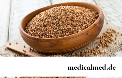
History of cultivation of a buckwheat contains more than five thousand years. Grain which is received from this plant is used for preparation of porridges, soups, baked puddings and puddings, do flour which is one of the main ingredients of the noodles popular in many countries of it. Buckwheat dishes are useful and tasty, they are perfectly combined with meat, milk, eggs, mushrooms, fruit and vegetables....
Section: Articles about health