Beam bone
Beam bone – the pair bone which is a part of a forearm and located knaruzh and kpered from an ulna.
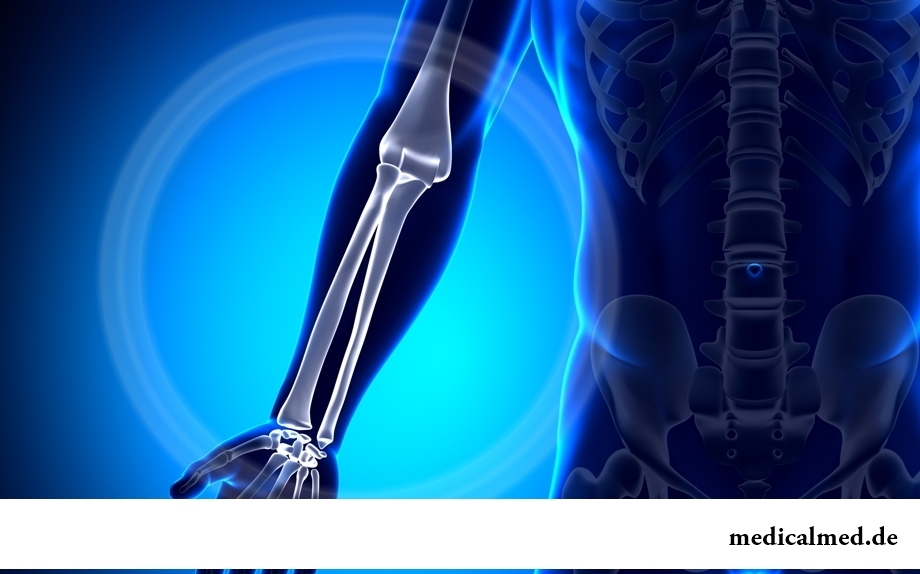
Structure of a beam bone
In a beam bone distinguish:
- Body of a trihedral form with three edges (front, interosseous and back) and three surfaces (front, back and lateral).
- Upper and lower epiphysis.
The front surface has a little concave form and in it the nutritious opening which the nutrient canal begins is located.
The smooth back surface is separated from a lateral surface by the rear edge.
Sinews of muscles lie in grooves of a back surface of the lower epiphysis of a beam bone.
Carpal joint surface call the place of a joint of the lower surface of a beam bone with wrist bones.
Fracture of a beam bone
Fracture of a beam bone in "the typical place" – the most often meeting among total number of various fractures of bones of a forearm. As a rule, falling arm-distance is the reason of this injury. At an injury in addition to a change there can be such accompanying damages as:
- Dislocation of a semi-lunar bone;
- Change of a navicular and awl-shaped shoot;
- Ruptures of sheaves (radiocarpal and radioulnar).
Most often elderly people, especially women are subject to changes that is connected with disturbance of exchange processes and development of osteoporosis.
Symptoms of a fracture of beam bone are:
- Sharp pain;
- "Forked" curvature of a forearm;
- Dysfunction of a brush and fingers;
- Hypostasis.
To establish whether there was a shift of a beam bone at a change it is necessary to make a x-ray film in two projections.
If necessary the traumatologist sets the displaced bone fragments then impose a plaster splint.
Control x-ray films, as a rule, carry out for 10-12 day after an injury (after fall of hypostasis). Sometimes after reduction of hypostasis the extremity is not fixed by rather qualitatively plaster splint that in an effect can lead to the secondary shift of fragments.
At a change without the shift of a beam bone the period of an immobilization (rest) makes from four to five weeks. If the change was followed by shift, gypsum is imposed up to eight weeks.
Posttraumatic dystrophy of a hand belongs to one of the main complications at the shift of a beam bone (differently – Turner's trophoneurosis). Can cause it too closely imposed plaster steak that happens, as a rule, because of increase of hypostasis for several days after an injury.
In certain cases at unstable fractures of a beam bone which tend to the secondary shift of fragments operation which will prevent development of deformation of a hand and a prelum of nerves is required. In some cases carry out an osteotomy with defect substitution by an artificial or bone. As a rule, the plate is deleted in seven months after function of a hand and a shape of a bone is completely recovered.
Rehabilitation after a fracture of a beam bone
Rehabilitation after a fracture of a beam bone is recommended to be begun as soon as possible (right after pain abates). It is desirable to carry out recovery complex which would include not only remedial gymnastics, but also massage, use of the warming ointments and compresses, physical therapy. Besides, for removal of loading some exercises are recommended to be carried out in warm water.
Exercises have to cover all free joints of the injured extremity. Especially it is necessary to pay attention to warm-up of fingers: they should be unclenched and squeezed, and also to collect various small objects (buttons, matches).
It is necessary to carry out all cycle of exercises throughout not less than half an hour on two-three times a day.
Process of recovery after a change is promoted by massage procedures using special ointments and gels:
- Activating a metabolism in a site of application;
- Removing an inflammation;
- Accelerating healing;
- Stopping pain.
The rehabilitation which is carried out in time after a fracture of a beam bone promotes not only to bystreyshy recovery of functionality of a hand, but also is prevention of development of a trophoneurosis.
The well-known drug "Viagra" was initially developed for treatment of an arterial hypertension.
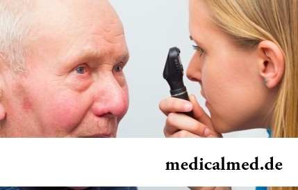
According to data of World Health Organization, the cataract is diagnosed almost for 7% of the population of Earth. Statistics we get sick...
Section: Articles about health
Smack in a mouth can arise in the natural way – as a result of lack of morning hygiene or reception of the corresponding food. However in certain cases its existence is a sign of certain pathologies, and allows to reveal an illness at an early stage. In we depend...
Section: Articles about health
The depression not without reason is considered one their main troubles of our century: for scientific and technical progress, acceleration of rate of life and a surplus of information of people it is forced to pay with stresses, negative emotions and weakening of protective forces of an organism. As a result widely the states which are characterized by the increased uneasiness, falling of interest in life, spiritual and physical discomfort extend....
Section: Articles about health
Quite large number of people adheres to the principles of vegetarian food. But how to be if in a family of vegetarians is д...
Section: Articles about health
About 10-15 years ago existence of the computer in the apartment of the Russian was considered as a rarity and office rooms were only at the first stage of equipment by these useful devices. Today practically in each house there is a computer (and often not one), and to constants...
Section: Articles about health
What is in our understanding weeds? It plants which are considered to be suitable only for compost pits and feeding of animals. Meanwhile, among the weeds growing literally under legs it is possible to find the mass of the officinal herbs having invaluable advantage for human health. It is possible not only to be treated by most of them, using as broths, tinctures, compresses, but also to accept in food as usual products. Let's consider 8 widespread and often ignored by people...
Section: Articles about health
The kid who was recently born is surrounded with love of adult family members and their cares without which the baby cannot exist....
Section: Articles about health
A lot of things depend on a condition of a backbone in a human body, a backbone - not only a support for a body, it also a receptacle for a spinal cord, that is why malfunctions with a backbone are so dangerous. To treat rachis diseases very difficult and long...
Section: Articles about health
The state of health of the person depends on many factors. One of the most important is the constant, but not exhausting a physical activity. In the presence of various illnesses specialists often advise patients to do swimming which by right takes the leading place by efficiency of improvement, having at the same time a few contraindications. Today we will talk about the main directions of therapeutic impact of swimming on a human body....
Section: Articles about health
Cold is such painful that each sigh becomes a victory, heat "knocks" down, and the ache in joints forces to think only about...
Section: Articles about health
The body of the person almost for 60% consists of water. It is so important for normal functioning of an organism that loss of only one and a half percent of liquid already leads to the most unpleasant effects. The problems connected with deficit of water can overtake and...
Section: Articles about health
The majority of gynecologic diseases prove three main signs, each of which speaks about need of a visit to the gynecologist. Certainly, it is possible to establish the exact diagnosis only after inspection, but on the basis of some signs it is possible to assume existence of this or that pathology. Let's consider symptoms of the female diseases which are found most often....
Section: Articles about health
Statistically, can only one of ten of our compatriots brag of a decent condition of an oral cavity. On среднестатистич...
Section: Articles about health
Today about 30 diseases, sexually transmitted are known. Wide circulation of these illnesses is extremely promoted by the dual attitude towards them: on the one hand, most of people know about "shameful" diseases and not a stirrup very little...
Section: Articles about health
The metabolism at each person proceeds in own way. However between the speed of this process and disposal of excess weight after all all have a dependence. Unfortunately, the people inclined to try on itself numerous "miracle" diets, not always consider this circumstance and with the most resolute intentions begin to eat so that artificially slow down the metabolism instead of to accelerate it. Except quite clear disappointment, incorrectly picked up systems...
Section: Articles about health
The modern person not always manages to find housing in the environmentally friendly region and such work which would not do harm здо...
Section: Articles about health
Any of us is not insured from a heavy illness of the loved one. Happens and so that someone from family members becomes the bed patient, and remains in such state for a long time. It extremely suppresses both the most injured, and all it to...
Section: Articles about health
High temperature - a frequent symptom of such widespread diseases as a SARS, quinsy, pneumonia, etc. To reduce heat, having facilitated a condition of the patient, doctors recommend to accept antipyretics, however their use is not always possible. Too frequent use of these drugs can lead to allergic reactions, and also overdose, causing poisoning. It happens also that there are no antipyretics simply in the house. In these situations it is pertinent to use it...
Section: Articles about health
The list of stereotypes of which, apparently, all know strongly includes following: British surely eat for breakfast овсянк...
Section: Articles about health
The dietology, as well as other sciences, does not stand still. Food stuffs are exposed to comprehensive study, and scientists obtain new information on their properties and influence on a human body. Unfortunately, this reasonable and natural process from time to time д...
Section: Articles about health
Coffee - the tonic loved by many for the invigorating aroma and deep taste. Having the stimulating effect, coffee increases working capacity, promotes concentration of attention, fights against drowsiness and improves mood. Statistically, about 30% of inhabitants of the planet regularly use coffee, from them more than 8% are "coffee-achievers" - the persons using more than 3 cups of drink a day....
Section: Articles about health
The fatigue, sleep debt, disturbances of food, bad mood, vagaries of the weather – all these circumstances badly are reflected in our vn...
Section: Articles about health
You are office worker, the driver, the fan of winter sports or do not think of life without bicycle? You lead a slow-moving life and you move on the city only on the car? You have no constant partner and you do not love the protected sex? Attention! You one...
Section: Articles about health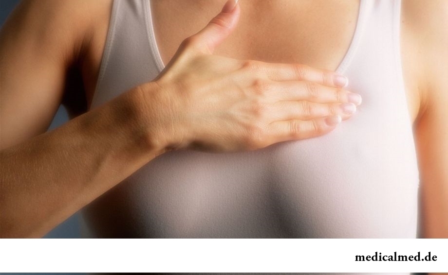
80% of women at least once to lives complained of discomfortable feelings to breasts, consolidations and nagrubaniye. These are mastopathy symptoms. The mastopathy is characterized by change of a ratio between ferruterous and connective tissue tissues of mammary glands. It can lead to formation of cysts (a cystous mastopathy), gland consolidation (a fibrous mastopathy), or a combination of these processes (a fibrous and cystous mastopathy)....
Section: Articles about health
(Xerostomia) many people consider feeling of a xerostomia small and easily removable inconvenience. This delusion...
Section: Articles about health
Since the moment when the child becomes a school student, his sight begins to be exposed to the strengthened loadings which are supplemented with viewing of animated films and long computer games. During this period of life of the child development not completely created bodies to a zra...
Section: Articles about health
Statistically, in Russia about 34% of citizens smoke. Most of consumers of tobacco has problems with health sooner or later. Not only smokers, but also their relatives suffer. Besides, cigarettes are expensive, and need of their acquisition goes a heavy burden on the budget of thousands of Russian families. Many people dream to refuse harmful tendency, but everyone manages to make it not: nicotine addiction is affectionate and to get rid of it not easy....
Section: Articles about health