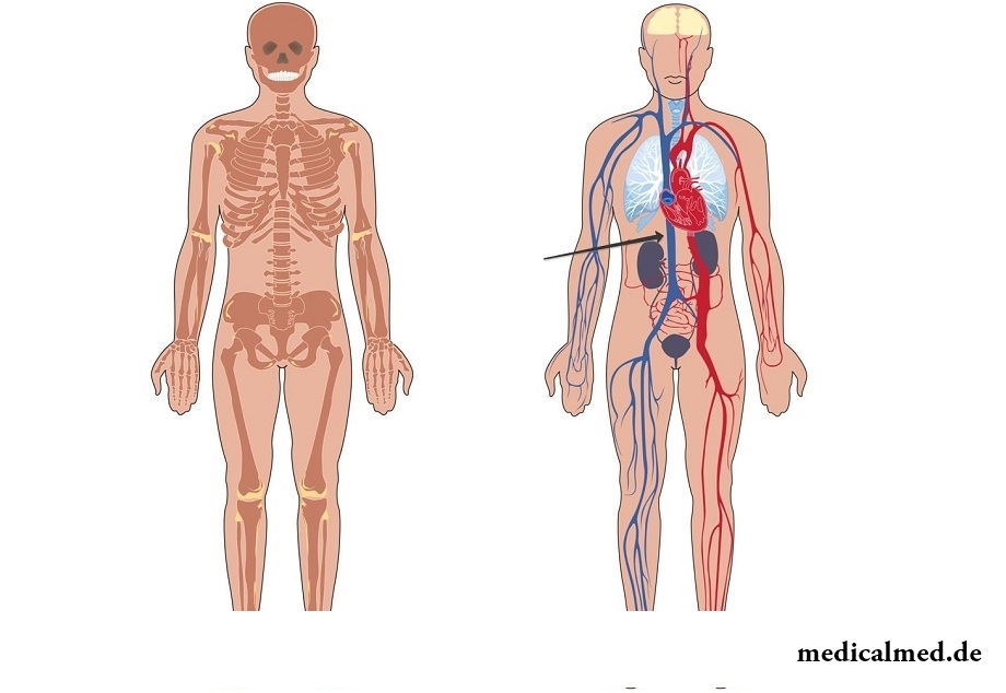
Lower vena cava
The lower vena cava – the wide vessel formed by merge of the left and right ileal veins at the level of the fourth or fifth vertebrae of lumbar department. Diameter of the lower vena cava varies ranging from 20 to 34 mm. Length of a chest part – 2-4 cm, belly 17-18 cm.
Structure of the lower vena cava
Vienna will be stirred in retroperitoneal space, behind internals, to the right of an aorta. It passes behind the upper site of a duodenum, behind a root of a mesentery and a head (top) of a pancreas and gets to a hepatic furrow, incorporating liver veins.
Passing through the opening of tendinous area of the same name of a diaphragm, the vein falls into the back area of a chest cavity. At the same time elastic, collagenic and muscle fibers of a wall of a vein are interwoven into a diaphragm wall.
Having reached a pericardium cavity, the vein gets into the right auricle. On the site of an entrance to the right auricle the vena cava is a little thickened. This vein has no valves.
Diameter of the lower vena cava changes during a respiratory cycle. At an exhalation the vein extends, and at a breath clenches. Change of diameter of the lower vena cava facilitates its recognition and differentiation from other large veins.
System of the lower vena cava
The system of the lower vena cava belongs to the most powerful system in a human body. About 70% of the general venous blood-groove fall to its share.
The system of the lower vena cava is created by the vessels collecting blood from an abdominal cavity, walls and bodies of a basin, the lower extremities.
This vein has parietal (pristenochny) and splanchnic (visceral) inflows.
Carry to pristenochny inflows:
- lumbar veins (on three-four from each party) – collect blood from muscles and skin of a back, from stomach walls, and also from area of a vertebral texture;
- phrenic veins – originate from the lower surface of a diaphragm;
- iliolumbar, lateral sacral, lower and upper buttock veins – collect blood from muscles of a stomach, a hip and a basin.
Carry to visceral inflows:
- gonadal veins – the ovarian and yaichkovy veins collecting blood from an ovary (small egg);
- renal veins - connect at the level of a cartilage to the lower vena cava between lumbar vertebrae (first and second). The left renal vein is much longer than the right renal vein. It crosses an aorta in front.
- veins of adrenal glands - the right vein gets into the lower vena cava, and the left vein connects to a renal vein.
- hepatic veins – bear blood from a liver.
All veins (except the largest) form numerous textures in and outside of bodies for blood redistribution. In case of injury of any vein the blood flow goes on collaterals (bypass ways).
Thrombosis of the lower vena cava
About 11% of total number of vein thromboses of a basin and the lower extremities are the share of thrombosis of the lower vena cava. The vein thrombosis can be primary and secondary (depending on the development reason).
Primary thrombosis develops owing to a malignant or benign tumor, inborn defects, a vein injury. The reasons of secondary thrombosis can be germination of a vein a tumor or its prelum. Quite often secondary thrombosis of the lower vena cava extends in the ascending way from other veins (smaller).
In medicine allocate thrombosis of the distal site of a vein, and also renal and hepatic sites. Thrombosis of the distal site of a vein is shown in cyanosis and hypostasis of the lower extremities, the lower half of a stomach, lumbar area. Sometimes hypostasis extends prior to the beginning of a thorax. The upper bound of cyanosis and a cutaneous dropsy depends on extent of spread of thrombosis.
At fibrinferment of a renal segment of a vein there are heavy general disturbances which can lead to a lethal outcome.
Development of thrombosis of a hepatic segment of a vein most often is followed by disturbance of the main functions of a liver and the subsequent thrombosis of a portal vein. Abdominal pains, increase in a spleen, liver, ascites, dispepsichesky frustration, change of a xanthopathy belong to symptoms of thrombosis of the hepatic site.
Prelum of the lower vena cava
The prelum of the lower vena cava can arise owing to a hyperadenosis, and also at retroperitoneal fibrosis and tumors of a liver.
The prelum of the lower vena cava and aorta the increased uterus at pregnant women serves as the reason of development of a syndrome of arterial hypotonia and emergence of disturbances of uteroplacental blood circulation (in a dorsal decubitus).
The vein prelum during pregnancy can lead to development of phlebitis, emergence of hypostasis of the lower extremities and venous stagnation.
There are very curious medical syndromes, for example, persuasive swallowing objects. In a stomach of one patient suffering from this mania 2500 foreign objects were revealed.

The fatigue, sleep debt, disturbances of food, bad mood, vagaries of the weather – all these circumstances badly are reflected in our vn...
Section: Articles about health
The saying "the rich do not know how the other half lives" is known to all. In a broad sense it is that we can not always understand the person whose features of a state are unknown to us. If with physiological characters of diseases the situation is more or less clearly (having noticed and...
Section: Articles about health
The phenomenon of the panic attack is known long ago, but the reasons of its emergence still are up to the end not found out. It is established that more than 30% of people at least once in life become the victims of very unpleasant phenomenon: without everyones on that the reasons they have a feeling of horror which is followed by a cardiopalmus, a shiver and the fever or feeling of sudden heat increased by sweating, breath constraint, dizziness, nausea....
Section: Articles about health
Modern footwear is extremely various. It stopped being only protection for legs long ago. Today shoes, boots, barefoot persons in...
Section: Articles about health
All parents are ready to what the baby often and pisat much. Since then, as the absorbing diapers strongly became current, keeping of the kid in dryness does not represent any problems. But if the grown-up kid continues to urinate in panties, parents of a nacha...
Section: Articles about health
Each of us repeatedly noticed that the people having the same passport age are sometimes not similar on one-years at all. One at the age of 40-45 years already looks almost an old man, and another and in 60 is young, vigorous and full of life. The matter is that the condition of our health depends not on the number of the lived years, and on degree of safety of an organism. This factor also defines biological age of the person....
Section: Articles about health
Producers of milk mixes for children assure: mixes are ideally balanced and adapted for needs of babies. In a sluch...
Section: Articles about health
An eye of the person daily experiences considerable strain. The problem of preservation of sight is for many years directly connected with a question of supply of tissues of eye enough oxygen and nutrients. This task is carried out by small vessels – capillaries. For holes...
Section: Articles about health
The next flu epidemic leads to the next panic, from year to year we give in on these manipulations: professionally alarming voice of the announcer in news, reports with calculation of the died patients, an interview with people in white dressing gowns and advertizing of anti-influenza means of different degree of inefficiency. All this reminds the Hollywood movies of epidemics threatening to destroy our planet. However, there is also one more similarity to cinema: everything comes to an end well. So, how to deal with the events, not in...
Section: Articles about health
Contrary to popular belief, the multiple sclerosis (MS) is not connected neither with sclerous changes of walls of vessels, nor about age...
Section: Articles about health
It seems, quite recently you brought the baby from maternity hospital, but time flew by, and here it is already going to join the first in life children's collective. How to prepare the child for visit of a garden? What needs to teach him to facilitate process адап...
Section: Articles about health
Mushrooms - the surprising inhabitants of our planet having a set of wonderful qualities. Thanks to one of them, a mold mushroom of Penicillium notatum, the first natural antibiotic - penicillin was received nearly 80 years ago. The mankind is obliged to this opening by millions of saved lives....
Section: Articles about health
No, probably, the person who would not have cold. Cold, cough, a headache – these symptoms are known to everyone. Peak to Prost...
Section: Articles about health
The body of the person almost for 60% consists of water. It is so important for normal functioning of an organism that loss of only one and a half percent of liquid already leads to the most unpleasant effects. The problems connected with deficit of water can overtake and...
Section: Articles about health
The words "disease" and "patient" not without reason come from one root – "pain". As a rule, symptoms of illnesses thoroughly spoil to patients life. However from this rule there are exceptions. Some diseases are shown by signs which can cause even positive emotions. It is a pity only that the majority of such illnesses are heavy and incurable....
Section: Articles about health
Childbirth is the most important event in life of each woman. We are women we give birth to the new little man on this light. Now...
Section: Articles about health
Household skills which to us so diligently imparted in the childhood it appears, not always bring only benefit. According to results of the last researches, some habits which for a long time were considered useful and even necessary can become...
Section: Articles about health
Practice of use of table salt in the therapeutic purposes contains not one century. Applications which do by means of the fabric impregnated with saline solution are considered especially effective. They have antibacterial and antiinflammatory effect, help to heal wounds, exempt fabrics from excess liquid. Hypertonic salt solution of potassium chloride is applied outwardly at many morbid conditions. Let's tell about the most known of them....
Section: Articles about health
All of us, unfortunately, should face flu nearly an every year. It would seem, so frequent disease has to be study...
Section: Articles about health
Cold is such painful that each sigh becomes a victory, heat "knocks" down, and the ache in joints forces to think only of pain. Some people with approach of the first symptoms of cold make the self-sacrificing decision to have a disease standing, and to a beam...
Section: Articles about health
Very often as a source of the infection which caused a disease serves our house - the place which a priori has to be safe. However disease-producing bacteria can perfectly feel not only in insanitary conditions, but also in our apartment if not to carry out due care of favourite places of their dwelling. What they − sources of their reproduction? Let's consider 10 most widespread places in our house, the most dangerous from the point of view of infection with microorganisms....
Section: Articles about health
We live during an advertizing era. Daily each person receives a solid portion of persuasive councils about what to eat to be здо...
Section: Articles about health
From the failure of work of immune system which is shown in the form of an allergy, statistically, more than 40% of the population of the globe suffer. In most cases pathological reactions cause the substances which are contained in food stuffs, hair of animals, medicines...
Section: Articles about health
What is in our understanding weeds? It plants which are considered to be suitable only for compost pits and feeding of animals. Meanwhile, among the weeds growing literally under legs it is possible to find the mass of the officinal herbs having invaluable advantage for human health. It is possible not only to be treated by most of them, using as broths, tinctures, compresses, but also to accept in food as usual products. Let's consider 8 widespread and often ignored by people...
Section: Articles about health
The advantage of swimming for the person is so high that this sport is not only the most popular, but also is widely applied in copper...
Section: Slideshow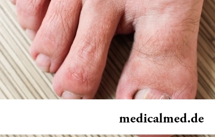
The word "onikhokriptoz" is unfamiliar to most of people, meanwhile quite so physicians call very widespread problem: the growing of edge of a nail into surrounding fabrics causing inflammatory process. Usually the illness affects thumbs of legs, and is followed покр...
Section: Articles about health
"Epilepsy" doctors made the diagnosis in antique times. Displays of an illness and pattern of its development are very well studied. However for nonspecialists this disease remains to not less mysterious, than in the ancient time. Many delusions are connected with epilepsy, and it sometimes very unpleasantly affects quality of life of patients and their relatives. In this article we will try to dispel the most known of similar myths....
Section: Articles about health