Eye retina dystrophy
General characteristic of a disease
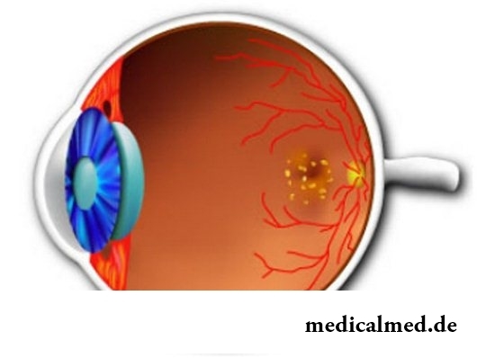
Eye retina dystrophy – one of the main reasons for poor eyesight of people of advanced age. The disease represents irreversible destruction of tissues of retina and gradual oppression of function of organs of sight.
The reasons causing eye retina dystrophy are happy are various. First of all, this age accumulation in fabrics of decomposition products of a metabolism. The major role in deficit of food of visual cells of a retina is played also by blood circulation disturbances, intoxication, infections.
The contributing factors to development of dystrophy of a retina of eyes is also short-sightedness. It leads to the supertension on eye covers caused by the increased cross size of organs of sight of the short-sighted person.
Presumably the diabetes mellitus, smoking, excess weight, high arterial pressure are considered as other factors starting the mechanism of development of dystrophy of a retina of an eye. Dystrophy of a retina of eyes can develop also at quite young age against the background of pregnancy, cardiovascular diseases, pathologies of a thyroid gland, and also as a result of injuries.
Retina dystrophy symptoms
Early symptom of dystrophy of a retina is gradual decrease in clearness of sight close. The disease slowly progresses and over time leads to perception of the distorted images. Patients with retina dystrophy symptoms quite often complain of doubling of visually perceived objects, a lomannost of lines, existence of blind spots at sight. However the total blindness as a result of dystrophy of a retina of an eye develops quite seldom.
Types of dystrophy of a retina of an eye
Chorioretinal dystrophy of a retina, it is an age macular degeneration of a retina, it is characteristic of people 50 years are more senior. The disease can lead to full loss of the central sight. At the same time peripheral sight of the patient with chorioretinal dystrophy of a retina remains within norm.
Without the central sight the clearness of perception of objects is impossible. People with symptoms of dystrophy of a retina of eyes of this type are not able to read or manage transport. Speed of progressing of a disease depends on type and weight of chorioretinal dystrophy of a retina.
The wet form of a disease is considered the heaviest, swift-flowing and badly giving in to treatment. However most often (in 9 of 10 cases) the dry form of chorioretinal dystrophy of a retina meets. At it process of gradual destruction of cells of the central part of a retina (makula) takes several years.
At other type of dystrophy of a retina of eyes, peripheral dystrophy of a retina, change mention peripheral departments of an eyeground. Danger of this disease is that the early stage of development of peripheral dystrophy of a retina proceeds almost asymptomatically. The first dystrophic changes can be diagnosed only by means of the special ophthalmologic equipment.
However it is very important to diagnose these changes at an early stage of peripheral dystrophy of a retina. Only in this case it is possible to warn effectively a serious complication of a disease - amotio or a rupture of a retina. Similar pathologies, unlike their prime cause, will quite hard respond to treatment.
Pigmental dystrophy of a retina – the most rare anomaly caused by hereditary factors. To it give disturbances in work of the photoreceptors of a retina answering or twilight black-and-white, or for day color vision.
Pigmental dystrophy of a retina can be transmitted from mother to the child on autosomal recessive or autosomal dominantly to a mode of inheritance. Most often men suffer from pigmental dystrophy of a retina. The clinical picture of a disease and the mechanism of development are deeply individual.
At pigmental dystrophy of a retina of easy degree there is only an insignificant decrease in visual acuity in badly lit space. In hard cases of pigmental dystrophy of a retina perhaps full fading of visual function.
Diagnosis of dystrophy of a retina of an eye
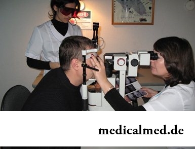
The most informative diagnostic methods of dystrophy of a retina of eyes are laser scanning of a retina by means of the optical tomograph, the central computer perimetry, and also a fluorescent angiography of blood vessels of an eyeground. They allow to reveal the earliest symptoms of dystrophy of a retina.
In addition at an early stage of diagnosis of a disease also tests can be applied to check of color sensation, contrast of sight, the sizes of the central and peripheral visual fields.
Treatment of dystrophy of a retina of an eye
In treatment of dystrophy of a retina of an eye of a chorioretinal form techniques of photodynamic therapy, laser photocoagulation, and also an injection of Anti-VEGF drugs are used. The last represent the special protein capable to suspend dystrophic processes in the makul of an eye.
Photodynamic treatment of dystrophy of a retina of eyes assumes intravenous administration of substances photosensitizers. They are capable to connect proteins of pathological vessels and to stop development of dystrophy. The mode of photodynamic treatment of dystrophy of a retina of eyes is set individually, depending on sensitivity of the patient to this type of therapy.
The technique of cauterization of pathological vessels is the cornerstone of laser treatment of dystrophy of a retina of eyes. On site a burn on fabrics of a makula of an eye the hem is formed, and sight on this site of an eye is not recovered. But this technique allows to prevent further distribution of process of dystrophy of a retina of an eye.
In treatment of pigmental dystrophy of a retina preferential physiotherapeutic techniques are used: magnito-and electrostimulation of tissues of eye. Unfortunately, their efficiency is not high. Also vazorekonstruktivny operations directed to improvement of blood supply of a retina have limited effect.
Prevention of peeling of a retina at peripheral dystrophy of a retina of eyes is carried out by means of laser coagulation. This low-invasive contactless technique of treatment of dystrophy of a retina allows to avoid surgical opening of an eyeglobe. The procedure is carried out on an outpatient basis and practically does not demand the recovery period.
As auxiliary treatment of dystrophy of a retina of eyes of all types also vitamin therapy is applied diyeto-.
It is known that with age contents in an organism of substances Luteinum and zeaxanthin, an eye, necessary for health, decreases. These substances are not produced in intestines therefore their contents needs to be filled regularly. Except zeaxanthin and Luteinum, the diet has to include vitamin C, tocopherol, selenium and zinc which feed, recover and protect tissues of eyes.
At complaints to manifestation of dystrophic changes of a retina and the progressing decrease in sight and, people after 45 years need to keep to a diet and to use means for sight support. For example, a vitamin and mineral complex of the European quality "Okuvayt Lyutein Forte" with Luteinum and zeaxanthin which protect eyes from a negative impact of a sunlight, C, E vitamins, zinc and selenium. It is proved that such structure prevents development of age changes of a retina of an eye, allows to enjoy acute eyesight even to elderly people.
Average life expectancy of lefthanders is less, than right-handed persons.

Life activity of one-celled fungi of the sort Candida is a proximate cause of development of candidiasis (milkwoman), it is related...
Section: Articles about health
The metabolism at each person proceeds in own way. However between the speed of this process and disposal of excess weight after all all have a dependence. Unfortunately, the people inclined to try on itself numerous "miracle" diets, not always at...
Section: Articles about health
Life does not indulge the modern woman special emotional comfort and carelessness. The fatigue, troubles at work, misunderstanding in a family and various illnesses immediately affect a condition of hair and skin. And there is a wish to look safe and attractive so! Substantially competently picked up diet can improve situation....
Section: Articles about health
Cold, puffiness of a nose, itch, the watering eyes - characteristic symptoms of the allergic rhinitis resulting from hit and...
Section: Articles about health
The number of long-livers is very small. One person from 5 thousand lives up to age of 90 years, and the centenary boundary steps over only one of 20 thousand. However, doctors claim that each of us is quite able to affect own destiny. At the same time speech to Ida...
Section: Articles about health
For the help to doctors in the choice of optimal solutions for treatment of various diseases the Cochrane scientific organization (Cochrane) conducts joint researches with representatives of scientific community around the world. The analysis of a series of the conducted researches of the drug Oscillococcinum® relating to group of cold remedies became one of the last methanolyses....
Section: Articles about health
Heart disease and blood vessels lead to disturbance of blood supply of bodies and fabrics that involves failures in their works...
Section: Articles about health
The state of health of the person depends on many factors. One of the most important is the constant, but not exhausting a physical activity. In the presence of various illnesses specialists often advise patients to do swimming which by right borrows ведущ...
Section: Articles about health
In consciousness of our many compatriots idea that folk remedies if are no more effective, than medicinal "chemistry" strongly took roots, then are precisely less harmful. Unfortunately, it is not always fair: some methods of treatment consecrated with "century national experience" can work so on the patient that it will need urgent intervention of physicians....
Section: Articles about health
Water with a lemon - idle time in preparation drink which supporters of a healthy lifestyle already managed to appreciate. Upo...
Section: Articles about health
Subfebrile temperature call fervescence to 38 degrees, and subfebrile condition - existence of such temperature over 3 days, and quite often it happens without the visible reasons. Existence of subfebrile condition - a strong indication of disturbances in an organism which can...
Section: Articles about health
Neurosis is called pathology of a nervous system at which deviations in functioning of the highest nervous processes are observed. Most often - owing to yet not strengthened mentality - children are subject to neurosises. The unhealthy, hostile atmosphere in collective, a family, the strong and sharp shock, and also a set of other factors which negatively influence the little person who did not learn to overcome stresses yet can become premises to emergence of such disturbances....
Section: Articles about health
Ability of an organism to resist to adverse environmental factors (to impact of temperature drops, humidity and pressure...
Section: Articles about health
Each of us faces from time to time that other people need the immediate help. We react to it differently: one at once call doctors and police, others rush to victims and try to save them independently. Some at all...
Section: Articles about health
Each person supports all life a SARS about 200 times. The peak of incidence falls on cold season, but it is possible to get sick with a temperature and a pharyngalgia, and sometimes and very possibly, even during a heat. The reasons for development of catarrhal diseases there is a set: from the weakened immunity till an excess portion of ice cream....
Section: Articles about health
The brain of the person is studied not one hundred years, but the quantity of the riddles connected with this body increases rather, than reducing...
Section: Articles about health
Dietary supplements (dietary supplements) for the last decades were so thoroughly included into our life that, apparently, it is already impossible to find the person who at least once did not try them. At the same time, most of our compatriots have a vague idea about...
Section: Articles about health
Weakness of an ankle joint – very widespread problem. Its existence is demonstrated by tendency to a podvorachivaniye of legs when walking on heels, frequent painful sprains, pain on average and anonymous toes even after small loadings. Usually people with such pathology take off unpleasant effects by means of the anesthetizing pulverizing and ointments, but it does not lead to radical elimination of a problem. Meanwhile, at the known persistence it is possible to strengthen an ankle to the house...
Section: Articles about health
One of the major chemical processes happening in a human body are oxidation reactions. They go with participation of fats...
Section: Articles about health
It seems, quite recently you brought the baby from maternity hospital, but time flew by, and here it is already going to join the first in life children's collective. How to prepare the child for visit of a garden? What needs to teach him to facilitate process адап...
Section: Articles about health
Venereal diseases in medicine are called the infections which are transmitted preferential sexually, now they and are called - infections, sexually transmitted, or STD. Among them is also life-threatening. In spite of the fact that the majority of diseases such will respond to treatment, they are widespread everywhere, and there is no tendency to decrease in incidence. Besides, some of them promptly look younger: statistically, a third of young people at the age of 16-22 years of a str...
Section: Articles about health
The popular expression "run from a heart attack" became the motto of the people supporting active lifestyle. Moreover, run became peculiar...
Section: Articles about health
New year, wedding, birthday, office party – an occasion to drink at the Russian person will always be. How to reduce a negative impact of alcohol by an organism and to avoid a condition of strong intoxication? The most correct council – to refuse the use spirits напитк...
Section: Articles about health
Use of medicinal plants in therapy is urgent today, more than ever. The drugs made of curative herbs cannot replace completely modern synthetic drugs, but their use becomes frequent serious help in simplification of a course of many illnesses and improvement of quality of life of chronic patients....
Section: Articles about health
EKO, or extracorporal fertilization - a method of treatment of infertility which became the reason of a set broken mines in due time...
Section: Articles about health
Deciding to get rid of an addiction, not all imagine what effects it is necessary to face. Process of refusal of smoking causes quite essential discomfort in most of people: differences of mood, sleep disorder, fatigue, decrease физич...
Section: Articles about health
The sudden heat on all body which is followed by perspiration and a cardiopalmus – the phenomenon familiar to many people. Most often such states called by "inflows" result from nervous or physical overworks and disappear right after rest. However in certain cases similar reaction of an organism can speak about diseases which need treatment. What? About it below....
Section: Articles about health