Fluorography of lungs
 Fluorography of lungs – a research of bodies of a thorax by means of the X-ray getting through pulmonary fabric and transferring to a film by means of fluorescent microscopic particles the drawing of lungs.
Conduct a similar research to the persons which reached 18 years. Frequency of its carrying out – is not more often than 1 time a year. This rule concerns only carrying out fluorography of healthy lungs when the additional examination is not required.
It is considered that fluorography of lungs – not rather informative inspection, but the data obtained with its help allow to reveal changes in a structure of pulmonary fabric and to become a reason for further more detailed inspection.
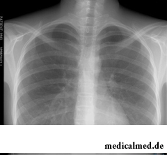
Bodies of a thorax differently absorb the radiation therefore the picture looks heterogeneous. Heart, bronchial tubes and bronchioles look light spots if lungs healthy, the fluorography displays pulmonary fabric homogeneous and uniform. And here if in lungs an inflammation, on fluorography, depending on the nature of changes of the inflamed fabric, are visible or blackouts – density of pulmonary fabric is increased, or the lightened sites will be noticed – lightness of fabric is rather high.
Fluorography of lungs of the smoker
It is established that changes in lungs and respiratory tracts imperceptibly happen even after the first smoked cigarette. Therefore to smokers – the people who are in a zone of the increased risk as regards pulmonary diseases it is strongly recommended to study fluorography of lungs annually.
 Not always the fluorography of lungs of the smoker will be able to show development of pathological process at its early stage – in most cases it begins not with lungs, and with a bronchial tree, but, nevertheless, the similar research allows to reveal tumors and consolidations in pulmonary fabric, the liquid which appeared in cavities of lungs, a thickening of walls of bronchial tubes.
It is difficult to revaluate importance of passing of such inspection by the smoker: the pneumonia which is timely found by means of fluorography, gives the chance to appoint as soon as possible necessary treatment and to avoid serious effects.
Interpretation of a flyuorogramma after passing of fluorography of lungs
Results of fluorography prepare usually several days, the flyuorogramma after that received is considered by the radiologist and in that case if the fluorography of healthy lungs was carried out, do not send for further inspection of the patient. Otherwise, if the radiologist found changes of pulmonary fabric, the person can be sent for specification of the diagnosis to a X-ray analysis or to an antitubercular clinic.
The conclusion of the radiologist in which such formulations can appear is attached to the picture received after fluorography of lungs:
- roots are expanded, condensed. Roots of lungs are created by lymph nodes and vessels, a pulmonary vein and an artery, a primary bronchus, bronchial arteries. Consolidation in this area at the general satisfactory condition of health indicates bronchitis, pneumonia and other inflammatory, perhaps chronic processes.
- Roots tyazhist. Most often such conclusion after the carried-out fluorography of lungs indicates bronchitis or other acute/chronic process. Such change of pulmonary fabric is often found on fluorography of lungs of the smoker.
- Strengthening of the vascular (pulmonary) drawing. The pulmonary drawing is formed by shadows of veins and arteries of lungs and if blood supply because of an inflammation is strengthened, and both bronchitis, and an initial stage of cancer, and pneumonia can do it, on fluorography it is noticeable that the vascular drawing is too selected. Besides, strengthening of the drawing revealed on fluorography of lungs can indicate also problems of cardiovascular system.
- Fibrous fabric. The found connecting fabric in lungs says that before people had a disease of lungs. It could be the injury, an infection or operation. In spite of the fact that the similar conclusion indicates loss of a part of pulmonary fabric, such result is often yielded by fluorography of healthy lungs.
- Focal shadows. So call blackouts of area of lungs on a flyuorogramma up to 1 cm in size. If the centers are found in the lower and average parts of lungs, it can be pneumonia. On a strong inflammation such formulation specifies in the conclusion of fluorography of lungs: "uneven edges", "merge of shadows", "strengthening of the vascular drawing". If more equal and dense, so inflammatory process declines the centers. If the centers are found in upper parts of lungs, it can indicate tuberculosis.
- Kaltsinata. So call the shadows, roundish in a form, reminding a bone tissue on density. The similar phenomena do not constitute danger, and only say that the patient had a contact with the patient with pneumonia, the tuberculosis infected with parasites, etc., but the organism did not allow to develop infections, and isolated bacteria activators under deposits of salts of calcium.
- Plevroapikalny stratifications, commissures. The structures found on fluorography of lungs from connecting fabric – commissures, in most cases also do not demand treatment, and only point to an inflammation in a pleura in the past. Sometimes commissures cause painful feelings, in this case it is necessary to ask for medical care. Plevroapikalny stratifications call thickenings of tops of lungs, and they also point that the person transferred the inflammation which affected a pleura (most often it is tuberculosis).
- The sine is soldered or free. Pleural sine are the cavities formed by pleural folds. If lungs healthy, the fluorography shows that sine are free. But liquid accumulation (in this case treatment is required) or the soldered commissures are sometimes observed.
- Changes of a diaphragm. Such conclusion after fluorography of lungs is drawn if anomaly of a diaphragm which could develop because of bad heredity, obesity, deformation by commissures, after postponed pleurisy is found in the person, diseases of a liver, gullet, intestines or stomach. In this case usually appoint additional inspection.
- The shadow of a mediastinum is displaced or expanded. A mediastinum call space between lungs and bodies in it being is an aorta, a gullet, heart, a trachea, absorbent vessels, nodes, a thymus. Expansion of a shadow of a mediastinum is observed because of increase in heart, a hypertension, heart failure, myocarditis. Shift of a mediastinum can indicate uneven accumulation of air or liquid in a pleura, big new growths in lungs. The similar conclusion of fluorography of lungs indicates that it is necessary to pass an additional examination and treatment immediately.
Our kidneys are capable to clear three liters of blood in one minute.

The fatigue, sleep debt, disturbances of food, bad mood, vagaries of the weather – all these circumstances badly are reflected in our vn...
Section: Articles about health
Separate food - the system of meal based on digestion physiology which is carried to improvement methods. According to nutritionists, the separate use of the carbohydrate and proteinaceous products demanding different conditions of assimilation helps to get rid from Bol...
Section: Articles about health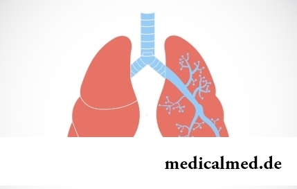
They say that to ensure health and longevity of people it is obliged. Really, at competent approach to these questions, minimization of an adverse effect of many factors does not represent a special problem. Practically everyone has an opportunity to play sports, to pick up an optimum operating mode and rest, to adjust healthy food, to refuse addictions. It is more difficult to exclude hit in an organism of harmful substances through a respiratory organs: not all are able to afford to live in the area with хо...
Section: Articles about health
Eyes – one of the most vulnerable areas on a face therefore age changes concern them first of all. Whether it is possible to keep a pier...
Section: Articles about health
There is a lot of fans of beer in our country. Statistically, on each average Russian (including women and children) in a year about 60 liters of this drink are consumed. It is not a lot of, as in the Czech Republic or Germany, but figure all the same impressive. Radova...
Section: Articles about health
Very often as a source of the infection which caused a disease serves our house - the place which a priori has to be safe. However disease-producing bacteria can perfectly feel not only in insanitary conditions, but also in our apartment if not to carry out due care of favourite places of their dwelling. What they − sources of their reproduction? Let's consider 10 most widespread places in our house, the most dangerous from the point of view of infection with microorganisms....
Section: Articles about health
The saying "the rich do not know how the other half lives" is known to all. In a broad sense it is that we can not always understand the person, about...
Section: Articles about health
Color of plants is caused by presence at them of certain chemical compounds. Let's talk about what is meant by various colors of vegetables and fruit and what properties they give them....
Section: Articles about health
Cold – a state known to everyone which is followed by cold, cough, high temperature, a pharyngalgia. Often the first that we begin to do in hope again to become healthy – to accept medicines which are not always harmless whereas it is easy to facilitate displays of a disease by means of natural means. They not only softly eliminate disease symptoms, but also enrich the weakened organism with useful substances. We present you 8 drinks which are successfully used for...
Section: Articles about health
New year, wedding, birthday, office party – an occasion to drink at the Russian person will always be. How to reduce a negative impact...
Section: Articles about health
The medicine promptly develops, and the fact that else quite recently it seemed by miracle can now. We are not surprised any more to the fact that people with artificial joints and extremities can play sports, organ transplantation became a routine, and the latest cancer medicine п...
Section: Articles about health
What woman does not dream of a beautiful and thick hair? While physicians developed difficult schemes on hair transplant, in the industry of hairdresser's art a few years ago there was a sensation – methods of hair extension appeared. It would seem, dreams came true: though the procedure of building also does not belong to the category cheap, practically any woman can increase several times the volume of hair, change their length and color – generally, to become the real beauty queen....
Section: Articles about health


Work of a brain is extremely complex and in many respects is not studied yet. It is confirmed also by the features of thought processes which are shown when the person sleeps. Let's tell about some of them....
Section: Articles about health
Summer in the heat. Many are going to spend vacation abroad. Travelers the tender seas, rest on beaches wait, for survey достоп...
Section: Articles about health
Aging — natural and inevitable process. Over time our skin loses elasticity, on it saggings are formed, the face form loses former clearness. The procedure of nitevy lifting (nitevy tightening) can successfully solve this problem. In order that it is better познако...
Section: Articles about health
Life of the modern woman is very difficult. Opportunities to realize itself are wide: it not only education and career, but also the most various hobbies from sport before needlework. It is not less important to build private life, paying an attention maximum to children, the husband, parents, friends. For all these affairs there is catastrophically not enough time therefore each of us tries to cut down as far as possible its expenses on necessary, but not the most fascinating housework. With it we are successfully helped by means...
Section: Articles about health
All parents are ready to what the baby often and pisat much. Since then, as the absorbing diapers strongly became current...
Section: Articles about health
80% of women at least once to lives complained of discomfortable feelings to breasts, consolidations and nagrubaniye. These are mastopathy symptoms. The mastopathy is characterized by change of a ratio between ferruterous and connective tissue tissues of mammary glands. It can bring...
Section: Articles about health
Turnip, radish, horse-radish – once these and other products enjoyed wide popularity at our ancestors, being not only the food sating an organism but also the medicines curing of many diseases. Unfortunately, the use of some of them got out of fashion long ago, and once favourite plants and vegetables almost ceased to make a contribution to human health. Inclusion of such products in a modern diet − an effective measure of prevention and treatment of diseases which seldom suffered...
Section: Articles about health
For the time being the perspective of heart diseases seems to most of people remote and foggy. But sooner or later практичес...
Section: Articles about health
About influence of fasting days on an organism it is told much – both about advantages, and about shortcomings. It is considered that fasting day in the form of a short-term monodiet is useful, promoting effective removal of slags from an organism whereas irregular, it is excessive п...
Section: Articles about health
The body of the person almost for 60% consists of water. It is so important for normal functioning of an organism that loss of only one and a half percent of liquid already leads to the most unpleasant effects. The problems connected with deficit of water can overtake also the healthiest person if he, for example, spends several hours under the scorching sun, without having taken with themselves drink, but is very simple to correct health in this case. It is much more difficult to minimize effects of other reasons about...
Section: Articles about health
The winter swimming in open reservoirs called in our country by "winter swimming" – officially recognized sport and one of the ek...
Section: Articles about health
Weakness of an ankle joint – very widespread problem. Its existence is demonstrated by tendency to a podvorachivaniye of legs when walking on heels, frequent painful sprains, pain on average and anonymous toes even after small nagruzo...
Section: Articles about health
Condition of lips (their morbidity, outward) – one of indicators of health of the person. The peeling, dryness, pallor, and also cracks in corners of a mouth can be not only the cosmetic shortcoming which arose owing to physical damages and weather conditions but also the satellite of some diseases and disturbances in an organism needing treatment. Let's consider 10 possible reasons of emergence of angular cracks (perleches) in corners of a mouth and ways of their elimination....
Section: Articles about health