Left ventricle
Left ventricle – one of four cameras of heart of the person in which the big circle of blood circulation providing a continuous blood flow in an organism begins.
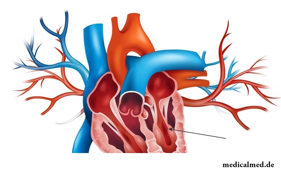
Structure and structure of a left ventricle
Being one of heart cameras, the left ventricle in relation to other departments of heart is located kzad, to the left and from top to bottom. Its outer edge roundish also carries the name of a pulmonary surface. The volume of a left ventricle in the course of life increases from 5,5-10 cm3 (at newborns) to 130-210 cm3 (by 18-25 years).
In comparison with a right ventricle left has more pronounced oblong and oval form and is slightly longer also than muskulisty.
In a structure of a left ventricle distinguish two departments:
- The back department which is a cavity of a ventricle and by means of the left venous opening is reported with a cavity of the corresponding auricle;
- The front department – an arterial cone (in the form of the output channel) is reported by an arterial opening with an aorta.
At the expense of a myocardium the wall of a left ventricle in thickness reaches 11-14 mm.
The internal surface of a wall of a left ventricle is covered with fleshy trabeculas (in the form of small ledges) which form network, intertwining among themselves. Trabeculas are less expressed, than in a right ventricle.
Functions of a left ventricle
The big circle of blood circulation which includes all branches, capillary network, and also veins of fabrics and bodies of all organism begins an aorta of a left ventricle of heart and serves for delivery of nutrients and oxygen.
Dysfunction and treatment of a left ventricle
Systolic dysfunction of a left ventricle call decrease in its ability to throw out blood in an aorta from the cavity. It is the most frequent reason of development of heart failure. Systolic dysfunction is caused, as a rule, by the contractility falling leading to decrease in its stroke output.
Diastolic dysfunction of a left ventricle call falling of its ability to pump over in the cavity blood from system of a pulmonary artery (otherwise – to provide diastolic filling). Diastolic dysfunction can lead to development pulmonary secondary venous and arterial hypertension which are shown as:
- Cough;
- Asthma;
- Paroxysmal night диспноэ.
Pathological changes and treatment of a left ventricle
The hypertrophy of a left ventricle belongs to one of typical damages of heart at an idiopathic hypertensia (differently – a cardiomyopathy). Development of a hypertrophy is provoked by changes in a left ventricle that leads to modification of a partition between the left and right ventricles and loss of its elasticity.
At the same time similar changes of a left ventricle are not a disease, and represent one of possible symptoms of development of any type of heart diseases.
Can be the cause of development of a hypertrophy of a left ventricle both an idiopathic hypertensia, and other factors, for example, heart diseases or considerable and frequent loadings. Development of changes of a left ventricle is sometimes noted for many years.
The hypertrophy can provoke the considerable modifications arising in the field of walls of a left ventricle. Along with a thickening of a wall there is a thickening of the partition located between ventricles.
Stenocardia is one of the most widespread signs of a hypertrophy of a left ventricle. As a result of development of pathology the muscle increases in sizes, there is a ciliary arrhythmia, and also are observed:
- Pain in a thorax;
- The increased arterial pressure;
- Headaches;
- Instability of pressure;
- Sleep disorders;
- Arrhythmia;
- Pain in heart;
- Feeling sick and general weakness.
Besides, similar changes of a left ventricle can be symptoms of such diseases as:
- Fluid lungs;
- Inborn heart disease;
- Myocardial infarction;
- Atherosclerosis;
- Heart failure;
- Acute glomerulonephritis.
Treatment of a left ventricle most often has medicamentous character along with observance of a diet and refusal of the available addictions. In certain cases the operative measure connected with removal of the site of a cardiac muscle which underwent a gipertrofirovannost can be required.
To small anomalies of heart, the shown existence in a cavity of ventricles tyazhy (additional connective tissue muscular educations) the false chord of a left ventricle belongs.
Unlike normal chords, false chords of a left ventricle have atypical fastening to an interventricular partition and free walls of ventricles.
Most often existence of a false chord of a left ventricle does not influence quality of life, but in case of their plurality, and also at an unprofitable arrangement they can cause:
- Serious violations of a rhythm;
- Decrease in portability of exercise stresses;
- Relaxation disturbances of a left ventricle.
In most cases treatment of a left ventricle is not required, however it is regularly necessary to be observed at the cardiologist and to carry out prevention of an infectious endocarditis.
One more frequent pathology is the heart left ventricular failure which is observed at a diffusion glomerulonephritis and aortal defects, and also against the background of the following diseases:
- Idiopathic hypertensia;
- Atherosclerotic cardiosclerosis;
- Syphilitic aortitis with defeat of coronal vessels;
- Myocardial infarction.
Insufficiency of a left ventricle can be shown both in an acute form, and in the form of gradually accruing circulatory unefficiency.
The main treatment at a left ventricular failure of heart is:
- High bed rest;
- Long inhalations by oxygen;
- Use of cardiovascular means – Cordiaminum, camphor, strophanthin, Corazolum, Korglykonum.
To tell even the shortest and simple words, we involve 72 muscles.

The advantage of swimming for the person is so high that this sport is not only the most popular, but also is widely applied in copper...
Section: Slideshow
All parents are ready to what the baby often and pisat much. Since then, as the absorbing diapers strongly became current, keeping of the kid in dryness does not represent any problems. But if the grown-up kid continues to urinate in panties, parents of a nacha...
Section: Articles about health
History of mankind contains several tens of epidemics whose emergence was compared by eyewitnesses and historians to doomsday. The most terrible of them claimed the lives of millions of people, having made even the whole people to the person of the earth. What they − the diseases striking terror? Whether it managed to the person to find treatment, or he is still powerless before forces of nature?...
Section: Articles about health
The hysteromyoma is diagnosed more than at a third of women 35 years are more senior. This high-quality new growth, which on early a stage...
Section: Articles about health
The climax, or menopause is the normal process of the termination of genital function of the woman which is followed by serious hormonal changes in an organism. Usually the menopause begins at the age of 50-55 years, but characteristics of this process are very individual. T...
Section: Articles about health
Nightmares belong to the most unpleasant frustration. Statistically, they happen at 4% of adults, and almost at 70% of children and teenagers. During a nightmare of people dreams himself in extremely difficult, life-threatening situation. He wakens suddenly, in a condition of a fright, and, as a rule, remembers the dream distinctly. The feeling of depression and alarm does not release throughout the day, creating hindrances for work and normal communication. If such episodes repeat often, can р...
Section: Articles about health
Tea is loved and use almost everything. This drink has tonic properties, contains the tannins capable podavlit...
Section: Articles about health
Small appetite at the child – the complaint which pediatricians should hear practically from each mother. Most often it is carried to the category of children's whims, however the refusal of food in certain cases can be to alarming symptoms therefore it cannot be ignored....
Section: Articles about health
More than a half of the married couples which faced prostatitis – leave. The new broadcast "Female View of Prostatitis" will help to learn – whether you have or your relatives problems....
Section: Articles about health
Sugar - the digestible refined product which is not of special value for an organism of the modern person. Use...
Section: Articles about health
Physical activity is necessary for normal functioning of a human body. At a lack of the movement joints cease to function, muscles atrophy, cardiovascular activity is broken and the metabolism worsens. Modern городс...
Section: Articles about health
Some people consider what for medicine of the 21st century of secrets in the field of health of the person almost does not exist. It absolutely not so. The more answers scientists receive, the more the most difficult questions are raised for them by life. Besides, there are diseases which are not explained with science in any way of which existence people know for 100-150 years. These diseases meet not so often, but from some of them nobody is insured....
Section: Articles about health
Household skills which to us so diligently imparted in the childhood it appears, not always bring only benefit. According to result...
Section: Articles about health
Diseases of joints often begin imperceptibly for the person. The first stages of destruction of the cartilaginous tissue providing soft and free sliding of heads of bones in joint bags proceed slowly and absolutely without serious consequences. Especially unpleasantly for that this пр...
Section: Articles about health
Contrary to popular belief, the multiple sclerosis (MS) is not connected neither with sclerous changes of walls of vessels, nor with age forgetfulness and problems with concentration of attention. This disease has the autoimmune nature. Pathological process is expressed in degradation of nervous tissue and destruction of its enveloping layer - a myelin. Multiple damages of the central nervous system which are shown by decrease in sight, bystry fatigue, on become result of development of an illness...
Section: Articles about health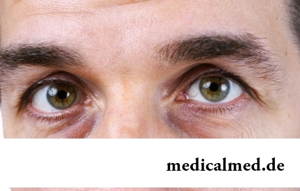
Zone hypostases under eyes - very widespread problem giving to people is a lot of inconvenience. Hypodermic fabric in these parts having...
Section: Articles about health
Phobia – the persuasive fear of a certain contents shown in a specific situation against the will of the person. Concepts of a phobia and fear are similar, however if the fear is natural protective function of mentality, then the phobia is its deviation. So the person can an ispytyva...
Section: Articles about health
Long time antibiotics were considered as a panacea from all diseases and were appointed even at insignificant symptoms of an infection. Even now not everyone knows in what force of antibiotics how and when they should be accepted. Let's discredit 7 popular myths about such drugs....
Section: Articles about health
Doctors claim that the people not so familiar with a dorsodynia occur among adult Russians very seldom. At the same time подавляющ...
Section: Articles about health
An eye of the person daily experiences considerable strain. The problem of preservation of sight is for many years directly connected with a question of supply of tissues of eye enough oxygen and nutrients. This task is carried out by small vessels – capillaries. For holes...
Section: Articles about health
Stroke (acute disorder of cerebral circulation) – one of the most widespread neurologic diseases. Annually in the world more than 6 million people die of this illness. From the survived patients about 80% become disabled people, and nearly a third from them needs afterwards permanent care. In fact, the stroke creates a situation at which a part of cells of a brain loses blood access, loses an opportunity to receive oxygen and nutrients, and perishes. As a result of a razviv...
Section: Articles about health
Cold, puffiness of a nose, itch, the watering eyes - characteristic symptoms of the allergic rhinitis resulting from hit and...
Section: Articles about health
Life of the modern child is extremely active and difficult. Information strain which is experienced by the school student and did not dream pupils of last times. Careful parents, wishing well to the children, will organize a set of additional classes in circles, sports...
Section: Articles about health
On the head of the person about one million hair follicles, or as they are called still, hair bulbs are located. At the time of the birth most of them is in the "sleeping" state, but within several weeks follicles become more active, and from them hair begin to grow. Intensity of this process is individual, and during life it can change. Genetic predisposition, a physical and emotional state, aggressive influence affects the growth rate of hair out of...
Section: Articles about health
Extracorporal fertilization – one of the most modern methods of controlling with infertility. So far it already helped znach...
Section: Articles about health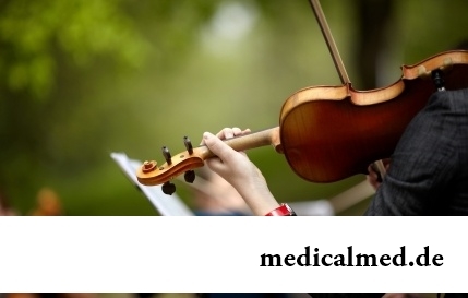
Musicotherapy – a treatment method which caused and causes a set of a controversy concerning its efficiency. However the facts are relentless: during the numerous researches curative impact of music on an organism was scientifically confirmed. Since then in a number of the countries a method...
Section: Articles about health
Each woman has preferences in the field of use of those goods which help us to look good, feel young and effective. Besides: selection process of favourite perfume, shampoo or decorative cosmetics already lightens the mood and serves as a peculiar stress medicine. Happens very offensively when the acquired perfumery and cosmetic products not only do not meet our expectations, but also becomes the reason of problems with health. Sources неприятн...
Section: Articles about health