


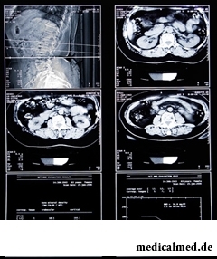
Tomography of an abdominal cavity
 By means of a tomography of an abdominal cavity the doctor obtains detailed information on a condition of the bodies which are in a peritoneum: kidneys, pancreas, liver, adrenal glands, spleen, vessels, abdominal lymph nodes. The computer tomography is successfully applied to diagnosing of diseases of blood: lymphogranulomatosis and lymphoma.
At men the tomography of abdominal organs helps to reveal ascites, inflammatory processes in a peritoneum, to define a prostate cancer, a rectum, bones, soft tissues, a bladder and its stage. Surely appoint a tomography for primary inspection at a basin injury, after operation or the carried-out rectum cancer radiotheraphy.
To women the tomography of an abdominal cavity is appointed for diagnosing of pathologies of a uterus, bladder cancer, at basin injuries, as primary inspection at inflammations in a basin.
The computer tomography is irreplaceable for diagnosis since as a result of its carrying out the quality three-dimensional image turns out, she allows to make cuts of internals the size 0,5mm and to reveal minor changes of bodies of a peritoneum which cannot be seen on other inspections.
Preparation for a tomography of an abdominal cavity
In the evening before the procedure the patient can eat only an easy dinner, in day of carrying out a tomography it is impossible to have breakfast. If inspection was appointed to the second half of day, only the light breakfast is allowed: liquid porridge, juice, coffee, firm food should not be.
 Besides, if use of contrast on inspection is supposed, the night before to the patient in the course of preparation for a tomography of an abdominal cavity appoint to drink small portions of half of liter of solution of Urografinum 15% (two ampoules of 75% of contrast solution part with 1,5 liters of boiled water). A half of liter more it is necessary to drink in day of inspection, in the morning and to take the remained 500 ml with itself and to drink in 30 and 15 min. prior to a tomography. It is possible to enter contrast intravenously – drug of Ultravist, 50-150ml.
Contrast when carrying out inspection is used if it is necessary to carry out diagnosis of cysts, tumors, a metastasis, pathologies of large vessels of an abdominal cavity. Contrast introduction directly on a tomography – is allowed when scanning of a certain interesting site.
The tomography of an abdominal cavity – is painless, during its carrying out the patient should lie not movably, periodically it is asked to hold for a while the breath. Prior to the procedure the patient needs to remove all objects containing metal. After the termination of a tomography it is possible to return to a usual way of life at once.
The tomography assumes radiation exposure, and, despite its low dose, the procedure with care, in urgent cases, is appointed to small children. In order that the child kept an immovability during the research, use of an anesthesia is in certain cases allowed. For the same reason apply anesthesia at inspection of seriously ill patients of patients.
Contraindications to carrying out a tomography of an abdominal cavity
Cannot appoint inspection the patient in whom the allergy to iodine and the drugs supporting him is found, at the increased function of a thyroid gland, heavy asthma bronchial, at the level of creatinine there is more 1,5mg/dl. The research is contraindicated to pregnant women, the feeding women.
Considering the available contraindications, to the patient before passing of a tomography appoint to take a blood test on biochemical analysis - for establishment of level of urea, ALT, nuclear heating plant, creatinine.
The well-known drug "Viagra" was initially developed for treatment of an arterial hypertension.

Nightmares belong to the most unpleasant frustration. Statistically, they happen at 4% of adults, and almost at 70% of children and...
Section: Articles about health
The naturopathy sometimes moves as the new direction of medicine, something like fashionable hobby, and there is nothing farther from the truth. This most ancient direction, the word "naturopathy" is translated as "treatment by the nature", and, no doubt, treatment приро...
Section: Articles about health
Food with the increased content of sugar is attractive to most of people - it is scientifically confirmed fact. Business here not in intemperance or dissoluteness: the sweet food is associated since childhood with feeling of rest and safety which tests the kid when it absorbs maternal milk. Besides, getting into a human body, sugar strengthens production of "happiness hormones" which all of us so need. And still life of sweet teeth seldom happens cloudless: their too big loss...
Section: Articles about health
Traveling all over the world, many try to try the most exotic dishes of national cuisines. Exists even so-called died away...
Section: Articles about health
They say that to ensure health and longevity of people it is obliged. Really, at competent approach to these questions, minimization of an adverse effect of many factors does not represent a special problem. Practically everyone has an opportunity to play sports...
Section: Articles about health
What woman does not dream of a beautiful and thick hair? While physicians developed difficult schemes on hair transplant, in the industry of hairdresser's art a few years ago there was a sensation – methods of hair extension appeared. It would seem, dreams came true: though the procedure of building also does not belong to the category cheap, practically any woman can increase several times the volume of hair, change their length and color – generally, to become the real beauty queen....
Section: Articles about health
What they, women? Beautiful, gentle, passionate and at the same time windy, gusty, and nervous. And what is stranger: all эт...
Section: Articles about health
About 10-15 years ago existence of the computer in the apartment of the Russian was considered as a rarity and office rooms were only at the first stage of equipment by these useful devices. Today practically in each house there is a computer (and often not one), and to constants...
Section: Articles about health
The summer of this year in Russia was very ambiguous. Regions suffered from a merciless heat, from pouring rains, the hail from time to time dropped out, then there was again a heat which alternated with rainfall again. Many people suffer from such sharp changes of weather. Even flu epidemics and a SARS were recorded....
Section: Articles about health
It would seem, about it there can be no disagreements: water is necessary for a human body for normal zhiznedeyatet...
Section: Articles about health
Cellulitis - very widespread cosmetic shortcoming which arises approximately at 80% of women sooner or later. Emergence it is connected with change of structure of a hypodermic fatty layer. At the same time on the surface of skin at first there are roughnesses (cambers...
Section: Articles about health
On the head of the person about one million hair follicles, or as they are called still, hair bulbs are located. At the time of the birth most of them is in the "sleeping" state, but within several weeks follicles become more active, and from them hair begin to grow. Intensity of this process is individual, and during life it can change. Genetic predisposition, a physical and emotional state, aggressive influence affects the growth rate of hair out of...
Section: Articles about health
When overcomes feeling of hunger, and an opportunity to have dinner fully is absent, having a snack − small on volume comes to the rescue...
Section: Articles about health
Osteoporosis this general disease which main sign is decrease in density of a bone tissue. On distribution width it takes the fourth place among noninfectious diseases. The illness develops at mature age more often: in our country to them harvest seasons...
Section: Articles about health
It is impossible to imagine human life in which there would be no plants. Practically in each apartment and any production room there are window plants, millions of people with pleasure are engaged in gardening and truck farming, many citizens spend free time on seasonal dachas. However we very seldom pay attention to those properties of our green pets who can make the neighbourhood with them unpleasant and even unsafe....
Section: Articles about health
The fatigue, sleep debt, disturbances of food, bad mood, vagaries of the weather – all these circumstances badly are reflected in our vn...
Section: Articles about health
All know that self-treatment is dangerous. However absolutely it is almost impossible to do without it. Rate of modern life does not allow to handle each small trouble to the doctor and information on ways of independent rendering medical the help...
Section: Articles about health
It would seem, to buy drugs in Moscow does not make a problem – a drugstore, and not one, is available for each resident of the capital within walking distance. And, nevertheless, Internet drugstores become more popular – what it is possible to explain such phenomenon with? Actually there is a lot of reasons and if to formulate them it is short, then the most suitable word will be - "conveniently". We suggest to get acquainted in more detail with pluses and minuses of online drugstores that buying drugs, not to make the wrong choice....
Section: Articles about health
We present to yours the TOP of the medicamentous means exerting the stimulating impact on a potentiality, i.e. on ability of a muzhcha...
Section: Articles about health
Each of us repeatedly noticed that the people having the same passport age are sometimes not similar on one-years at all. One at the age of 40-45 years already looks almost an old man, and another and in 60 is young, vigorous and full of life. The matter is that state нашег...
Section: Articles about health
Milk and products of its processing by right occupy one of the main places in a diet of the modern person. They contain proteins, necessary for normal life activity, fats, vitamins and microelements, and are an important part of various medical diets....
Section: Articles about health
Feeding by a breast - the integral part of ideal motherhood allowing to come into contact with the kid and to create since early years...
Section: Articles about health
Phobia – the persuasive fear of a certain contents shown in a specific situation against the will of the person. Concepts of a phobia and fear are similar, however if the fear is natural protective function of mentality, then the phobia is its deviation. So the person can an ispytyva...
Section: Articles about health
The majority of gynecologic diseases prove three main signs, each of which speaks about need of a visit to the gynecologist. Certainly, it is possible to establish the exact diagnosis only after inspection, but on the basis of some signs it is possible to assume existence of this or that pathology. Let's consider symptoms of the female diseases which are found most often....
Section: Articles about health
The number of long-livers is very small. One person from 5 thousand lives up to age of 90 years, and the centenary boundary steps only about...
Section: Articles about health
The sclera and mucous membrane of an eye are intensively supplied with blood vessels which problem - to saturate nervous tissues of body with nutrients and oxygen. In a normality vessels are almost not noticeable, however at their expansion (owing to истонч...
Section: Articles about health
Sometimes it seems that modern society was divided into two camps: representatives of the first are sure that only the woman has to be responsible for contraception, representatives of the second, respectively, are sure that it is destiny of men. Meanwhile the question of contraception has very many aspects – both psychological, and legal and, of course, medical....
Section: Articles about health