Backbone tomography
 Backbone tomography – detailed scanning and inspection of departments of a backbone.
Carry out a tomography computer and magnetic and resonant.
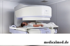
Computer tomography of a backbone
Data at a computer tomography are obtained by radiation of a backbone X-ray and the subsequent otsifrovaniye of the obtained data.
Preparing for inspection, the patient abstains from food – in 4 hours prior to a tomography of a backbone is it is impossible. Just before a tomography it is necessary to remove from himself metal objects (glasses, jewelry, prostheses, etc.). Those patients who are afraid of the closed space are recommended to accept soothing before inspection. At severe pains in spin it is possible to accept anesthetic.
During a backbone tomography the scanner of the tomograph moves around the patient and in the necessary foreshortenings does pictures.
After the first series of pictures is made, to the patient enter contrast and half an hour is watched reaction. If there are an itch, rash, there will be trouble breathing, the tomography of a backbone is not continued. If contrast did not cause an allergy, do the second series of pictures.
The result of the conducted examination represents pictures (tomograms) in which vertebrae are marked in white color, soft tissues – gray and cerebrospinal fluid – black. Besides, fabrics, various on density, also differ in the flowers.
The doctor recommends to make a backbone tomography for identification of the reasons of chronic and acute dorsodynias, assessment of a condition of a backbone before operation, diagnosing of cancer and a metastasis in intervertebral disks.
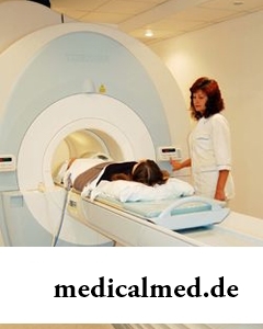
Magnetic and resonant tomography of a backbone
The magnetic and resonant tomography is considered safer for health since does not assume radiation: action of MR of the tomograph is based on the principle nuclear magnetic a resonance.
 MRT is considered the most exact, perfect and sensitive method of inspection of a backbone.
To make a backbone tomography by a method of magnetic resonance the doctor considers necessary for assessment of a condition of a backbone in general, for detection of a disease of vertebral fabrics, postoperative changes of a backbone, a condition of intervertebral disks, an inflammation and pinching of nerves, back infections, the reason of dorsodynias, tumors in a spinal cord or a backbone, for definition of date of operation on a backbone.
The patient for inspection is placed in the capsule, in it there is an electromagnetic field, and under its action molecules in a body of the person are located as well as electromagnetic waves. After that begin scanning of a backbone by means of radio waves. The backbone tomography lasts no more than 20 minutes.
The magnetic and resonant tomography of a backbone considerably surpasses computer since with its help it is possible to obtain more detailed information: the backbone is looked through in several planes better soft tissues are visualized.
Tomography of cervical and lumbar departments of a backbone
The magnetic and resonant diagnostic method at inspection of cervical and lumbosacral departments is irreplaceable. These sites are most subject to emergence of various defects and the majority of the conducted examinations of a backbone are the share of them.
Now MR a tomography of cervical department of a backbone, also as well as a tomography of lumbar department of a backbone are in the first place in comparison with a computer tomography, a X-ray analysis.
The tomography of cervical department of a backbone is appointed at osteochondrosis, hernia and a protrusion of disks, a stenosis of the channel of a backbone, metastasises of cells at the level of cervical department, injuries, anomalies of development of cervical department (a syndrome of additional cervical edges, a short neck).
Tomography of lumbar department of a backbone the doctor appoints MR to take place at osteochondrosis of lumbar department, hernia and a protrusion, tumoral metastasises, hemangiomas in vertebrae, an epidural space, a spinal cord, at a stenosis of the vertebral channel, injuries.
According to WHO researches the daily half-hour conversation by the mobile phone increases probability of development of a tumor of a brain by 40%.
The winter swimming in open reservoirs called in our country by "winter swimming" – officially recognized sport and one of the ek...
Section: Articles about health
Popular joke that there are no healthy people, and is nedoobsledovanny, most of us considers an honest truth, continually it is necessary to hear that all of us are sick hardly from a school bench. It is hard to say, whether so it actually because...
Section: Articles about health
History of cultivation of a buckwheat contains more than five thousand years. Grain which is received from this plant is used for preparation of porridges, soups, baked puddings and puddings, do flour which is one of the main ingredients of the noodles popular in many countries of it. Buckwheat dishes are useful and tasty, they are perfectly combined with meat, milk, eggs, mushrooms, fruit and vegetables....
Section: Articles about health
The modern person not always manages to find housing in the environmentally friendly region and such work which would not do harm здо...
Section: Articles about health
The medicine promptly develops, and the fact that else quite recently it seemed by miracle can now. We are not surprised any more to the fact that people with artificial joints and extremities can play sports, organ transplantation became a routine, and the latest cancer medicine п...
Section: Articles about health
Milk and products of its processing by right occupy one of the main places in a diet of the modern person. They contain proteins, necessary for normal life activity, fats, vitamins and microelements, and are an important part of various medical diets....
Section: Articles about health
Dietary supplements (dietary supplements) for the last decades were so thoroughly included into our life that, apparently, it is already impossible on...
Section: Articles about health
Ability of an organism to resist to adverse environmental factors (to impact of temperature drops, humidity and pressure, to the attacks of causative organisms, etc.) directly depends on what the person eats. Business here not only in, that C...
Section: Articles about health
Olive oil – the product capable to make a powerful contribution to health of the person if it includes it in the diet. The rich vitamin composition of oil does it by a product number one from many diseases including from deadly. Only two tablespoons of oil from olives in day prevent emergence of diseases of vessels and heart, cancer, problems with digestion, presenilation, a depression and many other illnesses which treatment would demand a lot of time and forces. Let's consider on...
Section: Articles about health
Partial and the more so full loss of hearing significantly reduces quality of life. Difficulties with communication lead to loneliness and замкн...
Section: Articles about health
So, you resolved to lose weight. And now you try to understand what to begin with: from exercise stresses or a diet? And how to make that process of weight loss did not give you an inconvenience, and, on the contrary, brought joy?...
Section: Slideshow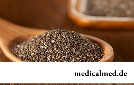
The chia plant, or the Spanish sage, is from South America. The indigenous people of the continent since ancient times used its seeds in food: small, but very nutritious kernels, in a form the reminding fasolina. Indians knew about useful properties of seeds of a chia, and applied them to maintenance of vitality and increase in endurance before serious exercise stresses....
Section: Articles about health


Venereal diseases in medicine are called the infections which are transmitted preferential sexually, now they and are called - infections, sexually transmitted, or STD. Among them is also life-threatening. In spite of the fact that the majority of diseases such will respond to treatment, they are widespread everywhere, and there is no tendency to decrease in incidence. Besides, some of them promptly look younger: statistically, a third of young people at the age of 16-22 years of a str...
Section: Articles about health
Obesity is called by a disease of 21 centuries, for the last 100 years by the number of the people suffering from excess body weight, considerably increased...
Section: Articles about health
Tea is loved and use almost everything. This drink has tonic properties, contains the tannins capable to suppress activity of causative organisms. Recently great popularity was gained by teas with vegetable additives. Лечеб...
Section: Articles about health
Run - one of the most available and effective ways to revitalize the organism. Knowing about its extraordinary advantage, each of us at least once tried to make jogs, but only the few made these occupations regular. In spite of the fact that in jogging (easy jogging), apparently, there is nothing difficult, the beginning runners often make mistakes which lead to complete cessation of trainings. Let's consider 10 useful tips for beginners who will allow them to make regular п...
Section: Articles about health
An eye of the person daily experiences considerable strain. The problem of preservation of sight is for many years directly connected with a question снабж...
Section: Articles about health
Smoking not only exerts a negative impact on the state of health of the consumer of tobacco products, but is an air polluter the substances potentially dangerous to people around. In recent years significantly the number of people, стремящ increased...
Section: Articles about health
According to doctors, more than a half of men of 25-50 years suffer from frustration of the urinogenital sphere, but the minority sees a doctor from them. And in vain - even the insignificant discomfort in the field of generative organs can serve as a symptom of an illness fraught with grave consequences for health. So - after 40 years - it is easy for most widespread disease of the sexual sphere of men to pass the first symptoms of prostatitis (weight in the bottom of a stomach, decrease in a libido), having written off for overfatigue and fatigue. Let's consider...
Section: Articles about health
Subfebrile temperature call fervescence to 38 degrees, and subfebrile condition - existence of such temperature from above...
Section: Articles about health
There is an opinion that at low temperatures safety of products is ensured longer and better thanks to what the refrigerator is considered the most suitable place for storage of food. In most cases it is fair, however there is a number of products, for a kotor...
Section: Articles about health
Many parents of children at the age of 2-4 years face excessively whimsical behavior of the child. The kid exhausts constant crying and whims not only the parents, but also himself. In what the reasons of children's whims. And how to fight with them?...
Section: Slideshow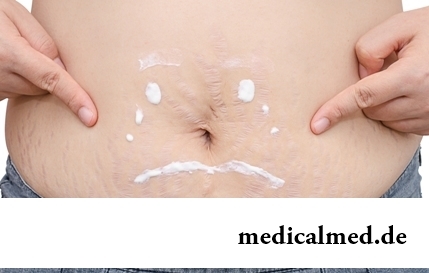
Striya (extension) are the defects of skin having an appearance of direct or wavy strips from 1 to 10 cm long and 1-5 mm wide. In the majority with...
Section: Articles about health
Cystitis, or inflammation of a mucous membrane of a bladder, this very widespread disease which, owing to some features of a structure of bodies of urinogenital system, women have approximately four times more often than men. In the main risk group...
Section: Articles about health
The metabolism at each person proceeds in own way. However between the speed of this process and disposal of excess weight after all all have a dependence. Unfortunately, the people inclined to try on itself numerous "miracle" diets, not always consider this circumstance and with the most resolute intentions begin to eat so that artificially slow down the metabolism instead of to accelerate it. Except quite clear disappointment, incorrectly picked up systems...
Section: Articles about health