Ultrasonography of a fruit
 Ultrasonography (ultrasonography) of a fruit is the main diagnostic method during pregnancy. How many time to do to the pregnant woman of ultrasonography of a fruit, the gynecologist, watching her defines. The purposes of performing ultrasonography of a fruit differ, depending on on what duration of gestation it is carried out.
Usually, the number of planned (obligatory) ultrasonography does not exceed 5 times:
1. For definition of the pregnancy – approximately on the term of 5 - 7 weeks;
2. For fetation assessment in a womb, and also a condition of a placenta of mother and an exception of malformations. Ultrasonography of heart of a fruit – on the term of 11 - 13 weeks is carried out;
3. For an exception of malformations, assessment of a condition of a placenta and amniotic liquid in it, and also for sex determination of the child. Surely the fruit sizes are determined by ultrasonography and ultrasonography of heart of a fruit – on term 19 – 21 week;
4. For determination of approximate weight of the child and a condition of an umbilical cord, and also commensurability of the size of his head and patrimonial ways of mother. The fruit sizes are determined by ultrasonography – on term 32 – 34 weeks;
5. For preparation for childbirth to expect possible complications – just before childbirth, with the first pains or at a bursting of waters.
Main types of ultrasonography of a fruit and as they are carried out
There is such ultrasonography main methods of a fruit as:
1. Transabdominal (the sensor is located on the woman's stomach);
2. Transvaginal (the sensor is entered into a vagina).
Both types of the procedure are absolutely painless for the woman, and ultrasonography is not harmful to a fruit.
Transvaginal ultrasonography is more exact method. Recently as very widespread ways of obtaining additional information on pregnancy are considered three-dimensional and four-dimensional ultrasonography of a fruit.
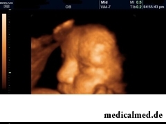 For three-dimensional or 3D-UZI the difficult computer program which provides the space image of a fruit which basis is made by two-dimensional (flat) pictures is used. In fact, three-dimensional ultrasonography allows to receive the exact photo of a fruit. Now such way of diagnosis is used for early detection of malformations of a fruit which could be missed during usual ultrasonography. For example, by means of ultrasonography of heart of a fruit it is possible to reveal defects in an organogeny.
Four-dimensional or 4D-UZI of a fruit allows to see the volume image of the child in real time, at the same time its movements and work of all internals are visible.
Future mothers are often disturbed by a question whether ultrasonography is harmful to a fruit. And so, modern devices ultrasonography are absolutely harmless to mother and future kid. Besides, they allow not only to determine the fruit sizes by ultrasonography, to make ultrasonography of heart of a fruit, but also to print out photos, and also to record video.
For performing ultrasonography of heart of a fruit the dopler of ultrasonography is used. This device gives the chance to conduct a blood circulation research in blood vessels, in heart of the child and in an umbilical cord, and also in vessels of a placenta of mother. Data a dopler of ultrasonography are important for identification at an early stage of potential problems with health of the child:
- Heart malformations;
- Anomalies of blood vessels;
- Problems with a placenta.
Dopler doctors recommend to carry out ultrasonography by everything to pregnant women on duration of gestation in 12 and (or) 20 weeks.
The gynecologist can appoint unplanned ultrasonography in the following cases:
1. Bloody allocations from a genital tract;
2. Pains in the bottom of a stomach.
Frequent repetition of ultrasonography for a fruit not harmfully and at all does not harm normal development of the child.
In order that it is correct to determine the fruit sizes by ultrasonography, to authentically estimate results of ultrasonography, having carried out interpretation of ultrasonography of a fruit, the pregnant woman needs to know the basic rules of preparation for this procedure. First, it is necessary to find out what type of ultrasonography is appointed (through a vagina or through a stomach). The way of preparation for ultrasonography depends on it:
1. When performing transabdominal ultrasonography approximately in 2 hours it is necessary to drink at least 1 liter of water and not to visit a toilet prior to a procedure;
2. When performing transvaginal ultrasonography the bladder had to be empty therefore, before the procedure it is necessary to descend in a toilet.
Besides, the woman before the procedure does not need to be nervous and ask a question whether ultrasonography is harmful to a fruit.
Interpretation of ultrasonography of a fruit
Norms of the indicators and parameters used for interpretation of ultrasonography of a fruit can vary, depending on duration of gestation. Interpretation of ultrasonography of a fruit is made by the doctor, by means of special tables.
The fruit sizes are determined by ultrasonography by the following indicators:
- Circle of a head of a fruit (HC);
- Biparietal diameter (BPD);
- Fruit length from a darkness to a sacrum (CRL);
- Length of a femur of a fruit (FL).
At interpretation of ultrasonography of a fruit the amount of amniotic liquid (amniotic waters) is defined. The aberration of this parameter, in the big or smaller party, can demonstrate disturbances of development of a nervous system or kidneys of a fruit, and also a signal of a pre-natal infection.
Much attention at interpretation of ultrasonography of a fruit is paid to a condition of a placenta (afterbirth). Ultrasonography determines the following parameters of a placenta:
1. Thickness;
2. Maturity degree;
3. Features of its attachment;
4. Condition of its development (for example, presentation).
Sex determination of the child by means of ultrasonography is done, usually, on the third planned ultrasonography (after the 20th week of pregnancy). The accuracy rate at sex determination by this method makes no more than 90%.
At interpretation of ultrasonography of a fruit there is possible a detection of the following anomalies of development:
- Hydrocephaly is accumulation in a head cavity of cerebrospinal fluid that threatens normal development of a brain;
- Anencephalia – total absence of a brain (the deadly diagnosis);
- The myelomeningocele is hernia of a spinal cord which seriously threatens development of a head and spinal cord of the child;
- Back of a bifid – process of splitting of a backbone. It threatens normal development of a spinal cord in the child;
- Fusion (atresia) of a duodenum – anomaly which demands urgent operation directly after the child's birth thanks to what it is possible to recover passability of intestines;
- Fruit malformations with ultrasonography of heart – deviations in its structure, than blood circulation in the child's heart is broken. It is important to reveal it that, in case of dangerous defect, to perform operation right after childbirth;
- The Down syndrome is a chromosomal disease at which multiple malformations and a delay of intellectual development of the child are observed.
Was considered earlier that yawning enriches an organism with oxygen. However this opinion was disproved. Scientists proved that yawning, the person cools a brain and improves its working capacity.

Household skills which to us so diligently imparted in the childhood it appears, not always bring only benefit. According to result...
Section: Articles about health
The modern person not always manages to find housing in the environmentally friendly region and such work which would not do harm to health. With food stuffs at first sight the situation is much better: shops are overflowed with goods which are positioned пр...
Section: Articles about health
Childbirth is the most important event in life of each woman. We are women we give birth to the new little man on this light. Now the tendency to that was outlined, as men want to participate in labor too. But there is a question and whether it is worth allowing the husbands on childbirth?...
Section: Articles about health
Condition of lips (their morbidity, outward) – one of indicators of health of the person. Peeling, dryness, pallor, and also трещ...
Section: Articles about health
There is an opinion that at low temperatures safety of products is ensured longer and better thanks to what the refrigerator is considered the most suitable place for storage of food. In most cases it is fair, however there is a number of products, for a kotor...
Section: Articles about health
Water with a lemon - idle time in preparation drink which supporters of a healthy lifestyle already managed to appreciate. Used in a warm look and on an empty stomach, it is one of the most useful prophylactics allowing to prevent tens of diseases and just to raise an organism tone. Especially effectively to use warm water with lemon juice after a serious illness, during a season of the colds, and also to children, old men and pregnant women which do not have contraindications...
Section: Articles about health
The climax, or menopause is the normal process of the termination of genital function of the woman which is followed serious hormonal...
Section: Articles about health
For many women the word "fat" sounds as a sentence. In aspiration to an ideal figure they try to exclude, first of all, from the menu all dishes containing fats without having at the same time a clear idea of a role of these substances in exchange processes, and about an afterbirth...
Section: Articles about health
Physical activity is necessary for normal functioning of a human body. At a lack of the movement joints cease to function, muscles atrophy, cardiovascular activity is broken and the metabolism worsens. The modern city rhythm of life does not provide the person with an adequate exercise stress, additional - sport is necessary. Tedious tasks the huge number of the people having those or ин exists sport not all to liking, but also...
Section: Articles about health
Healthy lifestyle today in fashion, and many parents think of that the child from the early childhood played sports. To a Torah...
Section: Articles about health
More than a half of the married couples which faced prostatitis – leave. The new broadcast "Female View of Prostatitis" will help to learn – whether you have or your relatives problems....
Section: Articles about health
Feeding by a breast - the integral part of ideal motherhood allowing to come into contact with the kid and to create to it healthy immunity since early years. Nevertheless, this important process in life of mother and child can be saddened laktostazy − by a milk delay in a mammary gland. What main reasons for a laktostaz? How not to allow problems with breastfeeding? Let's consider 10 premises resulting in stagnation of milk at the nursing mother....
Section: Articles about health
The name of this disease precisely reflects the problem reason: it consists in the bra fastener pressure upon a certain zone...
Section: Articles about health
Small appetite at the child – the complaint which pediatricians should hear practically from each mother. Most often it is carried to the category of children's whims, however the refusal of food in certain cases can be to alarming symptoms therefore it cannot be ignored....
Section: Articles about health
The main role in development of a peptic ulcer of a stomach and duodenum the bacterium Helikobakter plays pilor. Activity and the strengthened reproduction of this microorganism lead to weakening of protection of mucous membranes and their erosive damage. Displays of an illness seriously reduce quality of life: patients regularly test attacks of severe pain, heartburn, nausea. On this background also psychoemotional malfunctions develop: a kidney-vetch, as a rule, shows an acrimony, ча...
Section: Articles about health
The sudden heat on all body which is followed by perspiration and a cardiopalmus – the phenomenon familiar to many people. Most often t...
Section: Articles about health
The fatigue, sleep debt, disturbances of food, bad mood, vagaries of the weather – all these circumstances badly affect our appearance. Especially the person suffers: skin becomes flabby, loses healthy color, becomes covered by wrinkles, zones of hypostases and t appear...
Section: Articles about health
High temperature - a frequent symptom of such widespread diseases as a SARS, quinsy, pneumonia, etc. To reduce heat, having facilitated a condition of the patient, doctors recommend to accept antipyretics, however their use is not always possible. Too frequent use of these drugs can lead to allergic reactions, and also overdose, causing poisoning. It happens also that there are no antipyretics simply in the house. In these situations it is pertinent to use it...
Section: Articles about health
What woman does not dream of a beautiful and thick hair? So far physicians developed difficult schemes on hair transplant, in the bet industry...
Section: Articles about health
Each person knows that fervescence is an illness sign. However too low temperature (hypothermia), especially also can demonstrate existence of diseases when it is observed long enough. Such state is dangerous those...
Section: Articles about health
Phobia – the persuasive fear of a certain contents shown in a specific situation against the will of the person. Concepts of a phobia and fear are similar, however if the fear is natural protective function of mentality, then the phobia is its deviation. So the person can feel the unaccountable, baseless fear accompanied with neurotic symptoms (perspiration, a shiver, a fever) before any ordinary phenomenon – for example, a trip by the subway or a simple dog....
Section: Articles about health
The technique of acupuncture (acupuncture) is used in the medical purposes more than three and a half millennia. It widely races...
Section: Articles about health
Popular joke that there are no healthy people, and is nedoobsledovanny, most of us considers an honest truth, continually it is necessary to hear that all of us are sick hardly from a school bench. It is hard to say, whether so it actually because...
Section: Articles about health
Ability of an organism to resist to adverse environmental factors (to impact of temperature drops, humidity and pressure, to the attacks of causative organisms, etc.) directly depends on what the person eats. Business here not only in that cells of a body received a necessary set of nutrients, vitamins and microelements. Scientists established that such components which are capable to influence negatively immune system, in connection with also are a part of foodstuff...
Section: Articles about health
Some people consider what for medicine of the 21st century of secrets in the field of health of the person almost does not exist. It absolutely not so. Than Bol...
Section: Articles about health
Each of us faces from time to time that other people need the immediate help. We react to it differently: one at once call doctors and police, others rush to victims and try to save them independently. Some at all...
Section: Articles about health
Smack in a mouth can arise in the natural way – as a result of lack of morning hygiene or reception of the corresponding food. However in certain cases its existence is a sign of certain pathologies, and allows to reveal an illness at an early stage. Depending on character of aftertaste – acid, salty, bitter, sweet – distinguish also diseases which accompany it....
Section: Articles about health